
Page 80 - 2021_06-Haematologica-web
P. 80
J.C. Gutjahr et al.
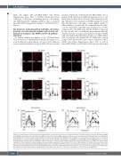
AML cells (Figure 4D) and OCI-AML3 cells (Online Supplementary Figure S6C) to VCAM-1 under shear flow conditions. Collectively, our findings point to a Src family- and PI3K-dependent signaling pathway that is initiated upon HA/CD44 engagement, leading to CD49d cluster for- mation.
The hyaluronic acid-induced VLA-4/VCAM-1 interaction promotes an acute myeloid leukemia cell-stromal cell interaction leading to Akt, MAPK and NF-kB pathway activation
To further identify the impact of the CD44-mediated VLA-4 activation on pathophysiologically relevant process- es in the BM microenvironment, we used a static adhesion assay combined with microscopy of AML cells on a stromal
cell layer. Untreated or HA-pretreated OCI-AML3 cells or primary AML cells from four different patients were co-cul- tured with stromal cells for 30 min. After extensive wash- ing, cell nuclei were visualized via DAPI staining. We found that HA-pretreated cells had a much higher ability to adhere firmly to stromal cells. We confirmed that this adhe- sion was dependent on the HA-induced interaction of VLA- 4 expressed by the AML cells and the VCAM-1 expressed by the stromal cell, as additional pretreatment with the αCD44 antibody as well as pretreatment with the αCD49d antibody inhibited the HA-induced adhesion of OCI-AML3 cells on stromal cells (Figure 5A, B). Additional controls using CD44 knockdown and control transduced OCI- AML3 cells (Online Supplementary Figure S7A) as well as native OCI-AML3 on αVCAM-1-antibody-treated stromal
Ci
Cii
Di Dii
Ai
Aii
Bi
Bii
Figure 4. Src family kinase inhibition and midostaurin treatment of acute myeloid leukemia cells inhibit hyaluronic acid-induced cluster formation. (A) Confocal images of OCI-AML3 (i) or primary acute myeloid leukemia (AML) cells from bone marrow (BM) aspirates (ii) that were or were not pretreated with HA and/or the Src family kinase (SFK) inhibitor PP2. Cells were stained with αCD49d (red) monoclonal antibody (clone AHP1225). For OCI-AML3, one representative experiment of three is shown; for primary samples, one representative experiment of six is shown (n=50 cells). (B) Confocal images of OCI-AML3 (i) or primary AML cells from BM aspirates (ii). Cells were pretreated or not with HA and/or the multikinase inhibitor midostaurin. Cells were stained with αCD49d (red) monoclonal antibody. For OCI-AML3, one representative experiment of three is shown; for primary samples, one representative experiment of six is shown (n=50 cells). (C) Mean numbers of CD49d clusters per cell were com- pared from six different patients’ samples with/without HA treatment and with/without PP2 treatment (i) or with/without midostaurin (ii). (D) Primary AML cells from five different patients with/without HA treatment and with/without PP2 treatment (i) or with/without midostaurin (ii) were subjected to shear flow analyses over VCAM-1. One- way analyses of variance with multiple comparisons were used. Bars, 5 mm. *P<0.05; **P<0.01; ***P<0.001; ****P<0.0001; ns: not significant.
2108
haematologica | 2021; 106(8)
