
Page 82 - 2021_06-Haematologica-web
P. 82
J.C. Gutjahr et al.
In conclusion, we demonstrate that HA-induced VLA-4 cluster formation is critical for direct cell-cell contact of human AML cells with stromal cells, thereby contributing to supportive signaling pathways in AML cells (Figure 7).
Discussion
The BM microenvironment plays a decisive role in the evolution and persistence of AML.1 Adhesive processes are mandatory to signal perception and leukemia cell-microen- vironment communication by facilitating the retention of the tumor cells to protective cues. Moreover, CD44 has been reported to be a marker of primary human AML can- cer stem cells and its blockade revealed a potential for dif- ferentiation in human AML cell lines.3,25,26 Here, we identi-
AB
fied a novel non-classical HA/CD44-triggered way of inside-out activation of the integrin VLA-4, leading to VLA- 4 cluster formation and increased adhesive strength on VCAM-1, important for the direct interaction of AML cells with supportive stromal cells.
In short-term adoptive transfers of human primary AML cells to immunodeficient mice, we observed that CD44 had a key function in rapid tumor cell homing to BM and spleen, reflecting the biology of normal cellular counter- parts as well as malignant cells, e.g. chronic myeloid leukemia-initiating cells.27,28 However, BM engraftment of malignant cells is dependent not only on homing events but even more on retention of the cells in distinct supportive zones of this organ.1 The VLA-4 integrin is known for being key for the retention of progenitor cells in BM.29 The strength of binding of the integrin to its ligand VCAM-1,
CD
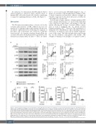
Figure 6. VLA-4 engagement triggers the phosphorylation of Akt, ERK, IκB and mTOR and contributes to acute myeloid leukemia progression. (A) Protein lysates of primary acute myeloid leukemia (AML) cells from bone marrow (BM) aspirates were treated or not with hyaluronic acid (HA) and/or VCAM-1-coated beads and tested for their ERK, phospho-ERK, Akt, phospho-Akt, IκBα, phospho-IκBα, mTOR and phospho-mTOR content by western blot. One representative experiment of five is shown. (B) Five independent experiments with five different AML patients’ samples were quantified. Expression intensities were quantified with ImageJ software and phospho- rylation was normalized to total protein content. One-way analyses of variance with multiple comparisons were used. (C) Apoptosis of native, shCont or shCD49d OCI- AML3 cells was induced with 0.5 mM of doxorubicin; additionally, cells were treated with immobilized HA and VCAM-1, where indicated. Cell viability was determined using trypan blue. Four replicates of one representative experiment of two independent experiments are shown. (D) Number of shCont or shCD49d OCI-AML3 cells in BM, blood, and spleen (SPL) of NSG mice, 28 days after intravenous injection (n=7 per group). *P<0.05; **P<0.01; ***P<0.001; ns: not significant.
2110
haematologica | 2021; 106(8)
