
Page 127 - 2021_06-Haematologica-web
P. 127
aGvHD impact on endothelium
ABC
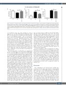
Figure 6. Reduction of endothelial apoptosis and endothelial activation by sildenafil in vitro. Mouse cardiac endothelial cells (MCEC) were incubated with either phosphate buffered saline (PBS)/ 0,1% dimethylsulfoxide (DMSO) (control [ctr]), 100 nm etoposide (eto), an inducer of cell death, or with 100 nm etoposide and 34 nm sildenafil (eto+sil) for 24 hours before analysis. (A) MTT assay showed higher optical density of eto+sil group versus eto-only group. (B) Staining for apoptotic cell marker caspase 3 (Casp3) showed reduced Casp3+ cells per high-power field (HPF) in eto+sil group compared to eto-only group. (C) Flow cytometry analysis of CD86, a costimulatory and endothelial activation marker, showed reduced percentage of endothelial CD86high cells in eto+sil group compared to eto-only group. Significance was tested by Student’s t-test (*P<0.05; n=2-3 experiments with at least triplicates per condition). Error bars indicate mean ± standard error of the mean.
at its initiation stage. As a first example for such an approach, we used the PDE5 inhibitor sildenafil, which has been demonstrated to normalize endothelial dysfunc- tion in vivo in different settings.32-39 First, we tested silde- nafil in vitro for EC protection from cytotoxic damage, mediated by etoposide. Sildenafil protected EC from etoposide-induced reduction of endothelial metabolic activity and proliferation (Figure 6A). Sildenafil signifi- cantly reduced etoposide-induced endothelial apoptosis, as quantified by Casp3+ staining (Figure 6B). For co-stim- ulatory capacity, we checked CD86high expression of EC by flow cytometry. Sildenafil treatment resulted in a sig- nificant reduction of etoposide-induced CD86 expression on EC (Figure 6C). Our data demonstrate a protective effect of sildenafil on etoposide-induced endothelial dys- function in vitro.
We tested the effect of sildenafil in our experimental models of aGvHD (Figure 7A to F) and SR-aGvHD (Figure 7G to L). In the aGvHD model without steroid treatment, we found that sildenafil treatment had no significant effect on survival (Figure 7A). However, sildenafil-treated allo- HSCT recipients with aGvHD had significantly lower clin- ical scores at different time points (Figure 7B) as well as lower histopathological aGvHD scores in the liver (Figure 7C) and colon (Figure 7D) as compared to untreated allo- HSCT recipients with aGvHD. In addition, we found a non-significant trend towards reduced costimulatory capacity and antigen presentation potential of hepatic EC under sildenafil treatment (Figure 7E and F). The vascular density (Online Supplementary Figure S9A) as well as the density of lymphatic vessels was not significantly affected by sildenafil treatment (Online Supplementary Figure S9B).
During SR-aGvHD, sildenafil treatment significantly improved survival (Figure 7G). Due to high mortality, clinical scoring was only significant in the early phase (Figure 7H). Histopathological aGvHD scores in the iver (Figure 7I) and colon (Figure 7J) were significantly reduced in sildenafil-treated allo-HSCT recipients with SR- aGvHD. Additionally, we found a trend towards reduced co-stimulatory capacity as well as significantly reduced antigen presentation potential of hepatic EC in sildenafil- treated SR-aGvHD, demonstrated by lower MHC class I and II expression (Figure 7K). We confirmed these find-
ings in another murine aGvHD model (129→B6, MHC- matched, Online Supplementary Figure S10). We found non-significant trends towards increased vascular density (Online Supplementary Figure S9C) and lymphatic vascular density in sildenafil-treated SR-GvHD versus controls (Online Supplementary Figure S9D). Next, we analyzed the effect of sildenafil on the endothelium during SR-aGvHD with electron microscopy analyses of liver and colon tis- sues (Figure 7M to P). We found endothelial damage in the liver and colon in untreated and sildenafil-treated SR- aGvHD. However, the sildenafil treated SR-aGvHD group showed reduced endothelial ruptures in liver (Figure 7N) and less prominent fibrinogen deposits in colonic biopsies (Figure 7P).
In order to analyze for effects of sildenafil on T-cell pro- liferation, we performed in vivo proliferation assays with carboxyfluorescein succinimidyl ester (CFSE) labeled allo- geneic T cells. In irradiated mice, sildenafil had no signif- icant effects on in vivo proliferation of allogeneic T cells (Online Supplementary Figure S11A to C). We next ana- lyzed and quantified the impact on sildenafil on immune cell subsets in peripheral blood during SR-aGvHD and found no significant differences (Online Supplementary Figure S11D to M). Our data suggest that the observed positive effect of sildenafil on SR-aGvHD is predominant- ly mediated by its effect on EC as opposed to effects on immune cells.
Discussion
The standardized and well-described aGvHD mice models provide the unique opportunity to experimentally address the role of endothelial dysfunction after HSCT. We found that severe endothelial damage, structural changes of the vasculature and endothelial dysfunction occur during aGvHD. Due to lack of suitable animal mod- els, less detailed data is available for other important endothelium-related complications of allo-HSCT, such as veno-occlusive disease and transplantation-associated microangiopathy. In line with the experimental results, we found that endothelial damage is present in the colon and duodenum of patients with severe aGvHD. This find-
haematologica | 2021; 106(8)
2155
