
Page 125 - 2021_06-Haematologica-web
P. 125
aGvHD impact on endothelium
considerable tissue damage in hematoxylin and eosin (H&E) staining is given in the Online Supplementary Figure S8, panels C and D. Patient characteristics and clinical information is given in the Online Supplementary Tables S4 to S6. Our data indicates that inflammatory activity in intestinal tissues is reduced after steroid treatment, while the endothelial damage is not influenced by steroid treat- ment during SR-aGvHD.
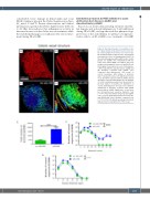
AB
Endothelial protection by PDE5 inhibition in acute graft-versus-host disease (aGvHD) and steroid-refractory aGvHD
Based on our results demonstrating extensive endothe- lial damage and reduced inflammatory T-cell infiltration during SR-aGvHD, we hypothesized that pharmacologic protection of the endothelium would have stronger pro- tective effects on SR-aGvHD versus treatment of aGvHD
Figure 4. Structural changes of vasculature in tar- get organs during acute graft-versus-host disease by scanning light sheet fluorescence microscopy. We analyzed organs at day+15 after experimental hematopoietic stem cell transplantation (HSCT) in the chemotherapy based B6→BDF model. Control groups (no aGvHD) were transplanted with the same bone marrow (BM) cell numbers and T-cell numbers from syngeneic donors. (A and B) VE-cad- herin signal of vasculature in colon of (A) allogene- ic-HSCT (allo-HSCT) recipients without aGvHD and (B) allo-HSCT recipients with aGvHD. (C-D) Computed three-dimensional (3D) model of colonic vasculature with number of branches (blue= low branch levels; red= high branch levels) of (C) allo-HSCT recipients without aGvHD and (D) allo-HSCT recipients with aGvHD. (E to G) Analysis of colonic vasculature parameters. Assessment of (E) total number of branches (F) distribution of vessels branching level and (G) vessel diameter distribution in allo-HSCT recipients with aGvHD versus without aGvHD. Significance of total num- ber of branches was tested by Student’s t-test (***P<0.001; n=3 animals per group) and signifi- cance of vessel branching level was tested by two- way ANOVA with Tukey’s multiple comparison test (***P<0.001; n=3 animals per group). Error bars indicate mean ± standard error of the mean.
CD
EF
G
haematologica | 2021; 106(8)
2153
