
Page 126 - 2021_06-Haematologica-web
P. 126
2154
S. Cordes et al.
haematologica | 2021; 106(8)
AB
CD
E
FGHIJ
KL
MNO
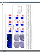
Figure 5. Inflammatory infiltration and endothelial damage in human steroid-refractory acute graft-versus-host disease (SR-aGvHD) and in an experimental model of SR-aGvHD. (A to E) Immune cell infiltration in colon samples at day+15 after experimental allogeneic hematopoietic stem cell transplantation (allo-HSCT) in the chemotherapy based B6→BDF model. Exemplary pictures of CD4+ infiltrates (green) in colon mucosa of (A) control phosphate buffered saline (PBS) treated (untreated) aGvHD and (B) Dexamethasone treated aGvHD (SR-aGvHD, 0.5mg/kg/day Dexamethasone starting at day+4). (C) Quantification of CD4+ area in colonic mucosa of untreated aGvHD versus SR-aGvHD. (D) Quantification of CD8+ area in colonic mucosa of untreated aGvHD versus SR-aGvHD. (E) Ratio of pericyte marker α smooth muscle actin (αSMA) positive area and endothelial cell marker CD31 positive area in in colonic mucosa of untreated aGvHD versus SR-aGvHD. Significance was tested by Student’s t-test (***P<0.001; n=5 animals per group). Error bars indicate mean ± standard error of the mean. (F to P) Human intestinal biopsies at aGvHD diagnosis and during the course of SR-aGvHD. (F to J) Intestinal biopsies at aGvHD diagnosis and SR-aGvHD stained against pan leukocyte marker CD45 (F) Colon biopsy stained against CD45 at aGvHD diagnosis with massive infiltrates. (G) Colon biopsy stained against CD45 during SR-aGvHD with low infiltrates. (H to J) Quantification of CD45 infiltrates at aGvHD diagnosis versus SR-aGvHD in (H) colon and (K) duodenum biopsies of cohort 1 and (J) in colon biopsies of cohort 2. (K-O) Intestinal biopsies at aGvHD diagnosis and SR-aGvHD stained against T-cell receptor marker CD3 (K) Colon biopsy stained against CD3 at aGvHD diagnosis with massive infiltrates. (L) Colon biopsy stained against CD3 during SR-aGvHD with low infiltrates. (M to O) Quantification of CD3 infiltrates at aGvHD diagnosis versus SR-aGvHD in (M) colon and (N) duodenum biopsies of cohort 1 and (O) in colon biopsies of cohort 2. (P) Quantification of capspase 3 positive (Casp3+) events in colonic endothelium of allo-HSCT recipients of cohort 2 at aGvHD diagnosis and during SR-aGvHD given in percent of vessels per high power field (HPF). Significance was tested by Student’s t- test (*P<0.05; **P<0.01; ***P<0.001; n= 4-6 patients per group). Error bars indicate mean ± standard error of the mean.
P
