
Page 129 - 2021_06-Haematologica-web
P. 129
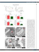
aGvHD impact on endothelium
GH
K
J
L
Figure 7. (G) Survival analysis and (H) clin- ical aGvHD manifestations of sildenafil (SR-aGvHD+sil) versus control substance treated (SR-aGvHD+ctr) SR-aGvHD. Histopathological assessment of aGvHD severity in (I) liver and (J) colon in SR- aGvHD+sil and SR-aGvHD+ctr at day+15 after allo-HSCT. Flow cytometry quantifi- cation of (K) MHCI and MHCII expression and (L) CD80 and CD86 expression of iso- lated liver sinusoidal endothelial cells of SR-aGvHD+sil and SR-aGvHD+ctr at day+15 after allo-HSCT. Significance was tested by Kaplan-Meier method and com- pared with the Mantel-Cox log-rank test (*P<0.05; n=8-10 animals per group) and Student’s t-test (*P<0.05; **P<0.01; n=6 animals per group). Error bars indi- cate mean ± standard error of the mean. Survival data was pooled from two experi- ments. All experiments were reproduced in a biological independent experiment and shown are representative results of one experiment. (M to P) Visualization of SR-aGvHD and sildenafil treated SR- aGvHD associated ultrastructural changes in the liver and the colon by transmission electron microscopy. Shown are typical pictures of sections from liver and colon taken at day+15 after experi- mental allo-HSCT in the chemotherapy based 129→B6 model. (M and N) Sinusoidal liver endothelial monolayer during SR-aGvHD and sildeanfil (sil) treat- ed SR-aGvHD. (M) Liver sinusoidal vessel of untreated SR-aGvHD with destroyed and unregularly shaped endothelial monolayer, marked by a red circle. (N) Liver sinusoidal vessel of Sildenafil treat- ed SR-aGvHD with small ruptures of the endothelial monolayer, marked by a red circle. (O and P) Colonic mucosa endothe- lium during SR-aGvHD and sildeanfil treated SR-aGvHD. (O) The vessel of untreated SR-aGvHD is surrounded by massive perivascular fibrinogen deposits marked by a red rhombus. (P) Occasionally little perivascular fibrinogen deposits, marked by a red rhombus, could be detected in sildenafil treated SR- aGvHD. V: vessel lumen; E: erythrocyte; N: nucleus; red rhombus: perivascular fib- rinogen deposits; red circle: loss of endothelium; red trapezoid: endothelial convolution).
MN
O P
haematologica | 2021; 106(8)
2157
I
