
Page 91 - 2021_07-Haematologica-web
P. 91
Fc-engineered CD54 antibody MSH-TP15 for myeloma therapy
AB
C
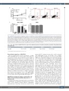
Figure 3. MSH-TP15 induces apoptosis of lymphoma cells after cross-linking on cell surface and inhibits myeloma growth in the presence of bone marrow stromal cells. (A) Induction of apoptosis was tested with Ramos lymphoma cells. After 6-hour incubation with the indicated antibody and 10 mg/mL anti-human fragment crys- tallizable γ (Fcγ) cross-linking antibody AV-FITC and 7-AAD positive cells were detected by flow cytometry. Rituximab (RTX) served as positive, a control immunoglobulin (Ig) G1 (ctrl IgG1) as negative control. Mean percentage ± standard error of the mean of four independent experiments is shown. (B) Results of one exemplified exper- iment with 10 mg/mL antibodies. AV-FITC-positive cells are marked in red boxes. (C) Growth inhibition of INA-6 myeloma cells was measured in the presence of bone marrow stromal cells (+BMSC) or absence of BMSC (-BMSC). MSH-TP15 and control IgG1 (ctrl IgG1) were used at 10 mg/mL (grey bars) or 100 mg/mL (black bars). Direct inhibitory effects on BMSC were not detected (100 mg/mL monoclonal antibody [mAb]; right graph). w/o: without; ctrl: control. Experiments were performed four times in triplicates. Graphs show mean values ± standard error of the mean. ***P<0.001, ** P<0.01.
Table 1. EC50 values achieved by MSH-TP15 Fc-Fc-engineered in antibody-dependent cell-mediated cytotoxicity experiments with peripheral blood mononuclear cells
Cell line INA-6
EC50 (nM) 0.36 (0.07-1.97)1
195% CI Confidence Interval, sigmoidal dose response curve.
Direct effector functions of MSH-TP15
First, we analyzed direct effector mechanisms of MSH- TP15. MSH-TP15 did not directly inhibit cell proliferation of myeloma cell lines (data not shown), but was capable of inducing apoptosis after antibody cross-linking on the cell surface. MSH-TP15 and rituximab both induced apoptosis in Ramos lymphoma cells expressing ICAM-1 and CD20, while a control IgG1 was not effective (Figure 3A). Total percentage of Annexin V (AV)-positive cells was calculated by combining early apoptotic (AV+/7-AAD–) and dead cells (AV+/7-AAD+) as shown for one exemplified experiment in Figure 3B. Next, we analyzed the impact of MSH-TP15 on growth of IL-6-dependent INA-6 myeloma cells in the pres- ence of BMSC isolated from myeloma patients. Growth inhibition of INA-6 cells in these co-culture experiments was observed in the presence of 10 or 100 μg/mL MSH- TP15, while no direct inhibitory effect on growth of BMSC was detectable with 100 μg/mL MSH-TP15 (Figure 3C).
MSH-TP15 Fc-engineered induces natural killer cell killing and macrophage-mediated phagocytosis of myeloma cells
In order to investigate Fc-mediated effector mechanisms of MSH-TP15, we performed chromium release assays. None of the antibody variants induced complement- dependent lysis of myeloma cells (data not shown), but sig-
nificant ADCC of plasmocytoma cells could be achieved with MSH-TP15 Fc-eng. and healthy donor’s PBMC (Figure 4A). Only minimal lysis was observed with MSH- TP15 and as expected no killing was detectable with the Fc k.o. variant and control mAb. EC50 values of MSH-TP15 Fc-eng. ranged from 0.36 nM (INA-6) to 3.77 nM (U266) and are summarized in Table 1. Of note, MSH-TP15 Fc- eng. also induced significant tumor cell lysis when patient- derived myeloma cells, including those from relapsed/refractory MM patients, and purified natural killer (NK) cells were used (Figure 4B, for patient details refer to Table 2). Flow cytometric analyses verified specific binding of MSH-TP15 to the CD138+ tumor cells of all patients (Figure 4B). NK cell induced killing of INA-6 myeloma cells by MSH-TP15 Fc-eng. was additionally investigated with live cell imaging (Figure 4C). Caspase activation and apoptosis of INA-6 cells was present over the entire 24 h assay time period. Exemplified microscopy images from 0, 6, 12 and 24 h are shown in Figure 4C. Quantification of the green fluorescent counts (caspase- dependent apoptosis) after 24 h revealed that significant killing of INA-6 myeloma cells occured only in the pres- ence of NK cells and MSH-TP15 Fc-eng. (Figure 4C). Finally, ADCP activity of the MSH-TP15 antibodies was analyzed with Raji lymphoma cells (CD19, CD20 and ICAM-1 positive) and monocyte-derived macrophages
L363 MM1.S U266
2.59 (0.87-7.77)1 1.82 (0.63-5.26)1 3.77 (0.83-17.2)1
haematologica | 2021; 106(7)
1861
