
Page 89 - 2021_07-Haematologica-web
P. 89
Fc-engineered CD54 antibody MSH-TP15 for myeloma therapy
AB
CDE
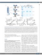
Figure 1. Generation and binding characteristics of MSH-TP15 antibody variants. (A) The variable heavy (VH) (dark blue) and variable light (VL) (light blue) sin-gle- chain fragment variable (scFv) sequences of phage antibody PIII-15 were used to generate three human immunoglobuin (Ig) G1 MSH-TP15 monoclonal antibody (mAb) variants carrying either a wild-type (middle), a protein-engineered (Fc-eng.; top) or a knockout (k.o.; bottom) fragment crystallizable (Fc)-domain. Mutations in the constant region (grey) of the heavy chain (HC) are depicted in yellow, glycosylation is shown in light grey. (B) Purity and molecular masses of the produced anti- bodies was analyzed by sodium dodecyl sulfate polyacrylamide-based discontinuous gel electrophoresis (SDS-PAGE) and Coomassie staining under reducing (red.) and non-reducing (n.r.) conditions. ADCC: antibody-dependent cell-mediated cytotoxicity; L: molecular mass ladder; LC: light chain. (C) Intercellular adhesion mole- cule-1 (ICAM-1) binding was measured by flow cytometry using L363 myeloma cells and increasing antibody concentrations. CD20 antibody rituximab served as con- trol (ctrl IgG1). ***P<0.001 MSH-TP15 mAb vs. ctrl IgG1. (D) Binding to human Fcγ receptor (FcγR) was analyzed with CHO cells expressing human FcγRIIa-131H or (E) BHK cells expressing either the low (FcγRIIIa-158F) or the high affinity (FcγRIIIa-158V) allelic form of human CD16a. *P<0.05 MSH-TP15/MSH-TP15 Fc-eng. vs. MSH-TP15 Fc k.o.; MFI: mean fluorescence intensity. Data represent mean values ± standard error of the mean of three independent experiments.
Data processing and statistical analyses
Data were statistically analyzed with GraphPad Prism Software using appropriate tests (San Diego, CA, USA). Significance was accepted with P<0.05.
Results
Generation of MSH-TP15 IgG1 monoclonal antibodies with different FcγR binding properties
VL and VH sequences of scFv-Fc antibody PIII-15, previ- ously selected by phage display and panning with human myeloma cell lines,22 were used to generate three fully human IgG1κ antibody variants. Compared to the wt IgG1 MSH-TP15, the Fc-optimized MSH-TP15 Fc-eng. and the Fc k.o. variant MSH-TP15 Fc k.o. were designed to display either enhanced FcγRIIa or FcγRIIIa binding to improve effector cell activation while retaining wt CDC activity or being incapable to mediate CDC and ADCC and therefore exclusively rely on Fab-mediated effector functions (Figure 1A). All proteins were produced in LentiX 293-T cells. Antibody preparations were highly pure and LC appeared at the calculated molecular masses of 25 kDa under reducing conditions. The slightly altered
migration of the calculated 50 kDa HC and 150 kDa IgG1 antibodies that were observed under reducing and non- reducing conditions is most likely due to glycosylation (Figure 1B). Importantly, the three MSH-TP15 mAb showed almost identical, concentration-dependent bind- ing to ICAM-1 with EC50 values in the low nanomolar (nM) range, but exerted significant differences in their FcγRIIa and FcγRIIIa binding (Figure 1C to E). As intend- ed, the MSH-TP15 Fc k.o. did not bind to these FcγR on stable transfected cells, while binding of MSH-TP15 Fc- eng. was significantly enhanced for both FcγR compared to wt MSH-TP15 IgG1 (Figure 1D and E). Thus, these results confirmed the expected differences of the three antibody variants.
MSH-TP15 binds domain 1 of human ICAM-1
In order to proof that by converting the scFv to a Fab fragment, epitope specificity was not altered, L363 cells were pre-incubated with an excess of TP-15-Fc (scFv-Fc antibody) prior to staining with Alexa Fluor 755-labeled MSH-TP15 IgG1. Importantly, MSH-TP15 binding was completely inhibited indicating the same ICAM-1 binding epitope (Figure 2A). In order to more precisely localize the binding region of MSH-TP15, truncated ICAM-1 variants
haematologica | 2021; 106(7)
1859
