
Page 81 - 2021_07-Haematologica-web
P. 81
anti-RhD antibodies modulate NK cell activity
RhD antibodies. In order to address this, we incubated KamRho with RhD+ erythrocytes and collected the super- natant, which consists of antibodies that did not bind to erythrocytes ("unbound" fraction, Figure 2A). As a control, we performed the same adsorption process with RhD- ery- throcytes, which are not expected to adsorb RhD-specific antibodies (Figure 1A). We first confirmed the efficiency of the process by staining RhD- and RhD+ erythrocytes with the two unbound antibody fractions. As expected, the unbound fraction from RhD- erythrocytes did not stain RhD- erythrocytes (Figure 2B, left panel), but did stain RhD+ erythrocytes (Figure 2B, right panel). In contrast, the unbound fraction from RhD+ erythrocytes no longer stained erythrocytes regardless of their RhD status (Figure 2C), thus demonstrating the efficiency of the adsorption process.
We next examined the effect of the unbound fraction on NK-cell degranulation. We found that the unbound fraction from RhD- erythrocytes highly activated NK cells, in con- trast to the unbound fraction from RhD+ (Figure 2D). In order to further corroborate the potency of specific anti- RhD antibodies in inducing NK-cell degranulation, we elut- ed the specific anti-RhD antibodies bound to RhD+ erythro- cytes ("bound" fraction, Figure 2E). As a control, we per- formed the same elution process with RhD- erythrocytes. We verified the elution process by staining RhD- and RhD+ erythrocytes with these bound fractions (Figure 2 F to G). Similar to the original antibody preparation, neither bound fraction stained RhD- erythrocytes (Figure 2 F to G, left pan- els). While the bound fraction from RhD- erythrocytes min- imally stained RhD+ erythrocytes, the bound fraction from RhD+ erythrocytes, as expected, significantly stained RhD+ erythrocytes (Figure 2 F to G, right panels). Both bound fractions were then incubated with NK cells and we deter- mined degranulation levels as before (Figure 2H). The bound fraction from RhD+ erythrocytes induced significant NK cell degranulation as compared to the bound fraction from RhD- erythrocytes (Figure 2H). Based on these results we concluded that it is the specific anti-RhD antibodies which induce NK-cell degranulation.
body. In concordance with our expectations, we observed binding of the anti-RhD antibodies to CD56dim NK cells that express CD16, but not to CD56bright CD16 negative NK cells (Figure 3D). The relatively low binding to CD56dim NK cells is probably due to limitations of staining human NK cells with human antibodies, as discussed below. Furthermore, blocking of CD16 on activated NK cells sig- nificantly reduced the binding of the bound RhD+ fraction to activated NK cells (Figure 3E).
In order to corroborate these results, we repeated the staining of freshly isolated NK cells with a fluorochrome- conjugated KamRho which we generated. This staining clearly demonstrated a specific binding to CD56dim (CD16+) NK cells (Online Supplementary Figure S2). Taken together, these findings indicate that anti-RhD antibodies bind CD16 on NK cells.
Next, in order to determine whether anti-RhD – CD16 interaction is responsible for the NK-cell degranulation, we repeated the degranulation assay with pre-blocking of CD16 on NK cells. Pre-blocking significantly reduced the degranulation levels induced by anti-RhD antibodies (KamRho in Figure 3F and Rhophylac in the Online Supplementary Figure S3A).
Anti-RhD antibodies induce natural killer cell degranulation by binding to CD16
B
We next explored how anti-RhD antibodies induce NK- cell degranulation. Because NK cells do not express the RhD antigen,16 we focused on CD16, the main FcR expressed by NK cells, as we suspected it binds the Fc portion of the anti- RhD antibodies. In order to test this possibility, we stained several transfected BW cells which express the extracellular domain of CD16 or of a control NK-cell receptor (NTB-A, NKp44 or DNAM-1). We first verified that each of the transfected BW cells expresses the extracellular domain of the given receptor (Figure 3A). We then stained these trans- fected BW cells with KamRho and analyzed the cells by flow cytometry (Figure 3B). We found that KamRho binds to CD16, but not to other tested NK-cell receptors (Figure 3B). Similar results were obtained with Rhophylac (Online Supplementary Figure S1C). Pre-blocking of CD16 with mouse IgG2a isotype, known to bind CD16 with high affinity,17 abolished the binding of KamRho to BW-CD16, as compared with IVIG binding (Figure 3C).
In order to further explore the possibility that anti-RhD
antibodies bind CD16, we double-stained primary freshly
+
isolated NK cells using the bound RhD fraction we gener-
ated (shown in Figure 2 E, G to H), and an anti-CD56 anti-
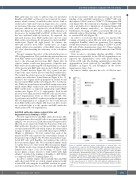
Figure 4. Anti-RhD antibodies induce degranulation of natural killer cells through their Fc segment. (A to B) Natural killer (NK)-cell degranulation assays.
A
In order to further elucidate the role of CD16 in anti-
fragments were produced from KamRho antibodies. NK cells were
2
(A) F(ab')
incubated with equal concentrations of the F(ab')
and intravenous immunoglobulin (IVIG). The NK-cell degranulation level was normalized to the basal percent of CD107a+ NK cells without antibody (no Ab.). (B) KamRho and IVIG were deglycosylated with PNGase under non-denaturing conditions. NK cells were incubated with equal concentrations of the deglycosy- lated or the original antibodies. The NK-cell degranulation level was normalized to the basal percent of CD107a+ NK cells without antibody (no Ab.). ***P<0.001; Student's t-test. Error bars represent standard deviation of tripli- cates.
2
fragments or whole KamRho
haematologica | 2021; 106(7)
1851
