
Page 82 - 2021_07-Haematologica-web
P. 82
S. Elias et al.
A
B
C
RhD–induced NK-cell degranulation, we cultured primary NK cells from donors who are homozygous for either of the low- or high-affinity alleles of CD16 (158F and 158V respectively).18 We performed degranulation assays with both NK cells in parallel, which were incubated with KamRho (Figure 3G) or Rhophylac (Online Supplementary Figure S3B). We found significant differences in the extent of NK-cell degranulation between the donors (Figure 3G; Online Supplementary Figure S3B). We therefore concluded that anti-RhD antibodies induce degranulation of NK cells by binding to CD16.
Fc glycosylations of anti-RhD antibodies are essential for inducing natural killer cell degranulation
As CD16 is an FcR, we wished to confirm that anti-RhD antibodies mediate their effect via their Fc segments. Therefore, we performed a degranulation assay with NK cells incubated with anti-RhD antibodies or F(ab')2 deriva- tives produced from KamRho and Rhophylac. As a control we used IVIG and F(ab')2 fragments of IVIG. In contrast to the degranulation induced by the whole anti-RhD antibod- ies, the F(ab')2 segments did not induce degranulation of NK cells (Figure 4A; Online Supplementary Figure S3C). This indicates that the Fc segment of anti-RhD antibodies is nec- essary for CD16-induced NK-cell degranulation.
In order to understand why the Fc fragments of anti-RhD antibodies, but not those of IVIG, activate NK cells, we investigated which element of the Fc is important for this effect. Since the human IgG subclasses 1, 2, 3, and 4 differ in their immune properties,19 we initially quantified the abundance of these subclasses in KamRho and Rhophylac, as well as in IVIG. Although some differences could be observed, we did not detect significant enrichment of a specific IgG subclass in the anti-RhD preparations as com- pared to IVIG (Online Supplementary Table S1), indicating that the functional difference is not due to a specific IgG subclass.
As Fc glycosylations have a critical influence on antibody
20,21
function, we next examined the role of Fc glycosylations
on anti-RhD-mediated NK-cell degranulation. We treated KamRho and Rhophylac with peptide:N-glycosidase F (PNGase F), an enzyme which cleaves N-linked oligosac- charides, and repeated the degranulation assay with the treated antibodies. Removal of glycosylations from KamRho and Rhophylac abolished their ability to induce degranulation of NK cells (Figure 4B; Online Supplementary Figure S3D, respectively), indicating that anti-RhD antibod- ies activate NK cells via glycosylation-dependent binding of their Fc segment to CD16.
Treatment with anti-RhD antibodies increases natural killer cell degranulation in humans
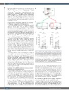
Since anti-RhD preparations are widely used in clinical settings we wished to determine whether anti-RhD anti- bodies affect NK-cell degranulation in humans. We there- fore collected peripheral blood samples from pregnant women who received prophylactic treatment with anti- RhD. We collected two samples from each woman: the first sample was obtained just before administration of the anti-RhD injection, and the second sample was obtained 3 hours post-injection. Samples were stained and analyzed for CD107a levels by flow cytometry with gating on NK cells (CD56+CD3-). Two NK-cell populations were observed and referred to as PBL1 and PBL2 (Figure 5A and B). We initially gated on the PBL2 population, which were
Figure 5. Treatment with anti-RhD antibodies increases natural killer cell degranulation in humans. (A-B) Gating strategy for assessment of natural killer (NK)-cell degranulation in human patients. Human peripheral blood (PB) samples were stained and analyzed by flow cytometry. (A) Size-based gating of two popu- lations of cells (PBL1 and PBL2). (B) NK cells were identified as CD3-CD56+ cells, after size-based gating. (C) Summary of the effect of anti-RhD treatment on NK cell degranulation in 11 women. NK-cell populations PBL2 (left panel) and PBL1 (right panel) were stained for CD107a. For each sample (before and after), NK-cell
+ degranulation was assessed by calculating the percentage of CD107a NK cells
out of the total NK cells. The triplicate average for each condition (before/after) of each patient was then plotted and used for the statistical test. Each black line represents a single patient. *P<0.05, Wilcoxon signed-ranks test.
larger in size and had high baseline degranulation levels, presumably representing activated NK cells. In accordance with the in vitro degranulation assays, we noted a signifi- cant increase in the degranulation levels in almost all patients (10 of 11) with an average 2.06-fold increase in NK-cell degranulation after receiving anti-RhD (Figure 5C, left panel). Similar results were observed when gating on the PBL1 NK population (Figure 5 A to C, right panels), with an average 1.34-fold increase in NK-cell degranulation after anti-RhD treatment (Figure 5C, right panel).
The anti-RhD drug KamRho induces killing of immature and mature dendritic cells by natural killer cells
Finally, we explored the possibility that anti-RhD-induced activation of NK cells helps to prevent production of anti- RhD antibodies by B cells, the desired clinical outcome of this treatment. As NK cells can kill iDC in peripheral tissues,
1852
haematologica | 2021; 106(7)
