
Page 80 - 2021_07-Haematologica-web
P. 80
S. Elias et al.
A
B
CDE
F G
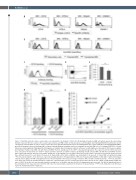
Figure 3. Anti-RhD antibodies induce natural killer cell degranulation by binding to CD16. (A to B) Staining of transfected BW cells which express the extracellular domain of specific NK-cell receptors. The receptor expressed by each BW cell line is indicated above each histogram. (A) Staining with different antibodies as indi- cated below each histogram, in order to verify receptor expression; gray filled histograms represent staining with isotype control antibodies. (B) Staining with KamRho; gray filled histograms represent staining with secondary antibody and gray histograms represent staining of the parental BW cells. (C) Staining of BW-CD16 cells with intravenous immunoglobulin (IVIG) (gray filled histograms) or KamRho (black histograms), without (left panel) or with (right panel) blocking of CD16 by mouse IgG2a. (D) Staining of freshly isolated NK cells with the bound RhD+ fraction. Gating was on the CD56+ cell population (gray box). The y-axis presents the CD56 expression level; cells above the black line are CD56bright (CD16-) and those below it are CD56dim (CD16+). (E) Staining of bulk activated NK cells with the bound RhD+ fraction, with or without blocking of CD16 by mouse IgG2a. Gating was on all living NK cells. The figure shows the average of three experiments, performed on NK cells from three different donors. For each donor, the percent of stained NK cells in both conditions was normalized to the percent of stained NK cells without blocking. *P<0.05; paired student’s t-test. Error bars represent standard deviation. (F) Degranulation of NK cells incubated with KamRho or IVIG, with or without blocking of CD16 by mouse IgG2a. The NK-cell degranulation level was normalized to the basal percentage of CD107a+ NK cells without antibody (no Ab.). **P<0.01; Student's t-test. Error bars represent standard deviation of triplicates. (G) Primary NK cells were isolated from donors who express the low- (158F) or high-affinity (158V) vari- ants of CD16. These NK cells were incubated with increasing doses of KamRho and then the NK-cell degranulation was assessed. The NK-cell degranulation level was normalized to the basal percent of CD107a+ NK cells. One representative experiment is shown out of two performed. ***P<0.001; Student's t-test. Error bars represent standard deviation of triplicates.
1850
haematologica | 2021; 106(7)
