
Page 182 - 2021_07-Haematologica-web
P. 182
A.G. Solimando et al.
AB
C
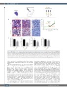
Figure 6. JAM-A inhibition restricts angiogenesis and tumor growth in subcutaneous multiple myeloma xenograft model. NOD/SCID mice (n=20) bearing RPMI-8226 subcutaneous xenografts were repetitiously treated with a JAM-A blocking monoclonal antibody (α-JAM-A), or isotope control IgG (ISO) or with vehicle only for 40 days for 3 days/week. (A) Immunohistochemistry staining: JAM-A (red) reactivity is stronger in the smaller neoplastic cells whereas it is lower in the more pleomorphic/anaplastic components. The opposite staining distribution is observed for Ki67 nuclear staining (brown), which is more clear-cut in the larger cells. CD31 staining shows focal pos- itivity in the control group and is absent in the group treated with the JAM-A blocking antibody. (B) Treatment was continued for 3 days/week for 40 days and tumor vol- umes were measured every 2 days with a caliper. (C) Hemoglobin values, Ki-67 positivity, vessel area and number of vessels expressed as mean ± standard deviation of three independent experiments. *P<0.05 versus vehicle-treated control. Scale bar=50 mm.****P<0.0001 versus controls; Mann-Whitney test.
effects, since JAM-A neutralization did not affect MMEC survival (Online Supplementary Figure S3A, left and right panels).
Based on the 2D observations, we investigated whether JAM-A could influence structured MM-associated angio- genesis in a 3D CAM assay. CAM were implanted with gelatin sponges imbued with either MMEC conditioned medium as the control (CTRL) or MMEC conditioned medium with sJAM-A (+sJAM-A), in the presence or absence of a monoclonal antibody blocking JAM-A. MMEC conditioned medium stimulated new vessel for- mation in CAM,19 and this effect was markedly enhanced by the addition of sJAM-A. The effect could be selectively inhibited by treatment with a sJAM-A blocking antibody as treatment with a cocktail containing an isotype IgG1 control antibody (sJAM-A + ISO, middle panel) did not reduce vessel formation (Figure 4C).
To explore potential factors that enhance JAM-A-medi- ated MM angiogenesis, we compared conditioned media
from MMEC supplemented with sJAM-A before and after treatment with the JAM-A blocking antibody with an angiogenesis array (Figure 4D, Online Supplementary Figure S3B). sJAM-A strongly reduced anti-angiogenic and increased pro-angiogenic factors secreted by MMEC,16 such as plasminogen (PLG), fibroblast growth factor 2 (FGF-2), insulin-like growth factor binding protein 1 (IGFBP1) and vascular endothelial growth factors A and C (VEGFA, VEGFC). Reverse transcriptase polymerase chain reaction analysis corroborated the proteomic findings and revealed sJAM-A-induced transcriptional upregulation of these factors and ligands (PLG and ENO1, JAM-A with LFA-1 and TJP1) (Online Supplementary Figure S3C-L). Moreover, because JAM-A can form homophilic interac- tions with JAM-A itself1 as well as heterophilic interac- tions with LFA-1, TJP1, CAV1 and CASK, we investigated whether the expression of these ligands correlated with MM-MMEC interactions. Direct co-culture of RPMI-8226 cells and MMEC significantly increased LFA-1 and CAV1
1952
haematologica | 2021; 106(7)
