
Page 178 - 2021_07-Haematologica-web
P. 178
A.G. Solimando et al.
A
B
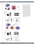
Figure 3. Bone marrow primary multiple myeloma endothelial cells enhance JAM-A expression on multiple myeloma cells. (A, B) Experimental design depicted at the top. RPMI-8226 cells were cultured alone or co- cultured with MMEC at a 1:5 ratio (RPMI- 8266:MMEC) in inserted transwells and ana- lyzed for JAM-A expression by western blotting (A) and flow cytometry (B). (C) Experimental design shown at top, RPMI-8226 cells were maintained for 24 h in CM from MMEC or MGEC. Cells were harvested and lysed and the extracted proteins immunoblotted for JAM-A expression. Overall densitometric analyses are reported. (D) RPMI-8226 cells were also analyzed by FACS after culture for 24 h in MGEC or MMEC CM. Results are pre- sented as mean ± standard deviation (MGEC from 24 patients with MGUS; MMEC from 24 patients with NDMM), ****P<0.0001, Mann- Whitney test. BM: bone marrow; MMEC: endothelial cells from patients with multiple myeloma; MGEC: endothelial cells derived from patients with monoclonal gammopathy of undetermined significance; NDMM: newly diagnosed MM; CM: conditioned medium; MFI: mean fluorescence intensity.
C
D
1948
haematologica | 2021; 106(7)
