
Page 54 - 2021_06-Haematologica-web
P. 54
C. Zhang et al.
AB
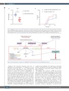
Figure 7. ALDH1A2 overexpression accelerates tumorigenesis in a zebrafish model. (A) Total reactive oxygen species levels in the mCherry-positive cells from the control and ALDH1A2-transgenic zebrafish were measured by flow cytometry and shown by mean fluorescent intensity as the fold-change compared to control cell samples. (B) Tumor development (lymphoma-like and leukemia-like phenotypes) in the single and double transgenic zebrafish was recorded according to the criteria defined by Langenau et al.50 Tumor onset and penetrance were evaluated by Kaplan-Meier curve analysis. *P<0.05 using the Gehan-Breslow-Wilcoxon test.
Figure 8. Scheme showing the oncorequisite role of ALDH1A2 for full transformation of T-cell acute lymphoblastic leukemia.
However, the molecular functions and roles of ALDH1A2 in the pathogenesis of T-ALL had not been elucidated previously. Here, we first experimentally proved that the T-ALL-specific isoform possesses an enzymatic activity that catalyzes retinaldehyde to retinoic acid with the production of NADH. Thus, the main mechanism involving this protein is characterized by ectopic expression through the activation of an alter- native promoter bound by TAL1 rather than the expres- sion of a dominant-negative protein or a loss of function. Additionally, we showed that this protein can reduce ROS levels and support energy production in T-ALL cells. Although the direct mechanism by which ALDH1A2 pro- motes the glycolysis pathway remains unelucidated, one possibility is that the high levels of NADH caused by ALDH1A2 overexpression might lead to pseudo-hypoxic
conditions that upregulate the expression of metabolic enzymes. Alternatively, the reduction in ROS level might lead to upregulation of metabolic enzymes. Further inves- tigation is needed.
In malignant cells, regulation of ROS and metabolic state is crucial to maintain cell proliferation and survival. Although it has been reported that ROS can promote the proliferation and survival of T-ALL cells,42 it is main- tained at low levels in the T-ALL leukemia-initiating cells in a mouse model.43 It is also noteworthy that, in more than 50% of the TAL1-positive T-ALL cases, genetic abnormalities in the PI3K-AKT pathway and the NOTCH1-MYC pathway have been observed,4,8-11,44 and these pathways can promote several metabolic process- es.45 In particular, MYC has been known to promote glu- taminolysis. Metabolic dependence on the TCA cycle
1556
haematologica | 2021; 106(6)
