
Page 52 - 2021_06-Haematologica-web
P. 52
C. Zhang et al.
ALDH1A2 overexpression accelerates tumorigenesis in a zebrafish model of T-cell acute lymphoblastic leukemia
Finally, we investigated the effect of ALDH1A2 overex- pression on tumorigenesis in vivo using a zebrafish model. We overexpressed the short isoform of the human ALDH1A2 gene with a fluorescent marker (mCherry) under the rag2 promoter in lymphocytes (Online Supplementary Figure S7A). We confirmed that transgenes were successfully integrated into the genome (Online Supplementary Figure S7B) and mCherry was expressed in the thymus (Online Supplementary Figure S7C). We then sorted mCherry-positive cells from the ALDH1A2-trans- genic and control fish and measured the ROS levels. Strikingly, the ROS level was significantly lower in the
ALDH1A2-transgenic fish than in the control fish (Figure 7A), supporting our results in cell lines.
We next monitored tumor development using off- springs from one of the established founder lines. However, we did not observe any spontaneous tumor development in the ALDH1A2 single transgenic fish (Online Supplementary Figure S7C). Hence, we then cross- bred this line with a transgenic line overexpressing a myristoylated, constitutively active mouse Akt2 gene (myr- mAkt2) which can cause T-ALL22 (Online Supplementary Figure S7B). Interestingly, overall penetrance was signifi- cantly increased up to 60% and tumor onset was slightly accelerated in the double transgenic animals as compared to the myr-mAkt2 single transgenic animals (Figure 7B,
A
B
CD
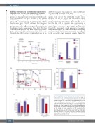
Figure 5. ALDH1A2 supports cellular aerobic glycolysis and energy production in T-cell acute lymphoblastic leukemia cells. (A) Real- time curves showing the normalized extracellular acidification rate (ECAR) readings for the control (blue) and ALDH1A2-depleted (red) cell samples. Bar graphs with normalized ECAR showing the changes in glycolysis, glycolytic capacity and glycolytic reserve after ALDH1A2 depletion. (B) Real-time curves showing the normalized oxygen con- sumption rate (OCR) readings for the control (blue) and ALDH1A2- depleted (red) cell samples. Bar graphs with normalized OCR show- ing the changes in basal respiration and ATP production after ALDH1A2 depletion. (C) Quantification of the NADH to NAD+ ratio after ALDH1A2 depletion. (D) Quantification of the ATP levels after ALDH1A2 depletion. All error bars represent the standard deviation for technical replicates. **P<0.01, ***P<0.001, ****P<0.0001 using the two-tailed Student t test.
1554
haematologica | 2021; 106(6)
