
Page 153 - 2021_06-Haematologica-web
P. 153
Effect of G-CSF on mouse HSC
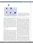
Figure 6. Cytokine network at the early stage of hematopoiesis. The model shows the cytokine network among HSC1, HSC2, HPC1, HPC2, HPC3, and HPC4. HSC1 cells respond to stem cell factor (SCF) and thrombopoietin (TPO). HSC2 cells respond to TPO and granulocyte colony-stimulating factor (G-CSF). HPC1 and HPC2 cells respond to SCF, TPO, and G-CSF. HPC3 cells respond to SCF, TPO, G-CSF, and GM-CSF. HPC4 cells respond to G-CSF and GM-CSF.
data suggested that HSC1 cells were quiescent in the steady state. After culture, most cells were continuously cycling in SCF + TPO, while approximately half of the cells were cycling in SCF or SCF + G-CSF.
Cell cycle progression is regulated by Cdkn1a (p21), Cdkn1b (p27), and Cdk1c (p57).26 p21 was not expressed in the majority of freshly isolated Mki67– cells (Figure 5) but p21 was expressed in most Mki67+ cells after culture; in particular, in SCF + TPO, its relative expression level was significantly increased. Interestingly, p27 was expressed in most freshly isolated Mki67– cells as well as most Mki67+ cells after culture. p57 was expressed in some of freshly isolated Mki67– cells, and also in some Mki67+ cells after culture; however, its relative expression level was decreased after culture. These data suggested that p21 was expressed in cycling cells while p57 was expressed in some quiescent cells. p27 was expressed in both quiescent cells and cycling cells.
Discussion
Functionally distinct HSC have been classified into My- bi, Bala, and Ly-bi HSC; α, b, and γ cells; or ST- and LT- HSC by different criteria. However, these classified cells overlap one another.22 Particularly, My-bi HSC overlap LT- HSC, and Ly-bi HSC overlap ST-HSC. In this study, by definition, we detected LT-My-bi HSC and ST-Ly-bi HSC at the single-cell level. We used HSC1 and HSC2 cells as highly purified HSC. HSC1 cells are significantly enriched in LT-My-bi HSC, while HSC2 cells are significantly enriched in ST-Ly-bi HSC.16,18-21 As shown in clonal trans- plantation, however, a small proportion of HSC1 con- tained ST-Ly-bi HSC (Figure 4A). Therefore, ST-Ly-bi HSC co-existed with LT-My-bi HSC in HSC1. We used HPC1, HPC2, HPC3, and HPC4 as highly purified HPC. We have recently shown that LT-HSC can be similarly detected in HSC1 and HPC1 cells.21 However, this study showed that Csf2rb expression in HPC1 was significantly greater than in HSC1 (Figure 1C), and HPC1 but not HSC1 cells responded to G-CSF in single-cell culture (Figure 2A).
HSC1 and HPC1 as populations remained functionally distinct.
Hematopoietic cytokines play a critical role in the regu- lation of hematopoiesis. In this study, we examined the effects of SCF, TPO, G-CSF, and GM-CSF on HSC1, HSC2, HPC1, HPC2, HPC3, and HPC4. SCF/c-Kit signaling plays an important role in hematopoiesis, particularly in the interaction of HSC and their niche, as shown by studies of W and Steel mutant mice.27,28 It has recently been reported that SCF is a niche factor from endothelial cells and perivascular stromal cells to maintain HSC.29 Li and Johnson were the first to report that SCF is a survival fac- tor of HSC in culture. We now confirmed their finding by transplantation assays (Figures 3 and 4). SCF alone was sufficient to support the survival of LT-HSC. Clonal trans- plantation assay showed that LT-HSC activity was detect- ed in progeny from single LT-HSC. Therefore, self-renew- al division took place in SCF culture (Figure 4B and Online Supplementary Table S6). Interestingly, HSC stopped divid- ing after 1-2 times in SCF culture (Figure 2). Single-cell RT- PCR suggested that some cells may be non-cycling, based on the expression of Mki67 (Figure 5). The role of p57 in regulating HSC quiescence has been suggested.30,31 However, we showed that p57 was not upregulated in Mki67– cells by SCF. p21 was expressed in the continuous- ly cycling cells in Mki67+ cells from SCF + TPO culture, but not Mki67– cells from freshly isolated cells. p21 expres- sion was upregulated in Mki67– cells after SCF culture, compared to Mki67– cells from freshly isolated cells (Figure 5). These 'stop-dividing' cells may differ from the quies- cent state in vivo at the molecular level. It would be inter- esting to see the functional difference between G0 and pseudo-G0 HSC.
Mpl was the second highly expressed receptor in HSC and HPC (Figure 1C). SCF and TPO synergistically acted on HSC1, HSC2, HPC1, HPC2, and HPC3, but they did not exert much action on HPC4 because the percentage of division and colony sizes in HPC4 were significantly smaller than those in the others (Figure 2B). Consistent with our data, it has been reported that deletion of TPO did not affect the number of CD34+FLT3+KSL cells but sig-
haematologica | 2021; 106(6)
1655
