
Page 151 - 2021_06-Haematologica-web
P. 151
Effect of G-CSF on mouse HSC
ative expression level of Csf3r also increased after culture with SCF + TPO but not with SCF + G-CSF. Suppressor of cytokine signaling (SOCS) family are physiological reg- ulators of several cytokine signaling.23 Less than 50% of HSC1 expressed SOCS3 while most HSC1 expressed SOCS 2-6, approximately 30% of HSC1 expressed SOCS1, and <30% of HSC1 expressed SOCS7. Interestingly, the expression of SOCS3, but not the other SOCS, increased after culture with SCF + G-CSF or TPO (Online Supplementary Figure S4A and B) suggesting a posi- tive correlation between Csf3r and SOCS3 expression in HSC. Members of early growth response gene (Egr) fami- ly, Egr2 and Egr3, can directly induce SOCS3 expression.24 Egr3, but neither Egr1 nor Egr2, was detected in most cells (Online Supplementary Figure S4A and B). A very small num- ber of freshly isolated HSC1 cells expressed Cxcr4, and the percentage of Cxcr4-expressing cells did not increase after culture with SCF, SCF + G-CSF, and SCF + TPO. These
data suggested that these cytokines cannot directly upreg- ulate the expression of Cxcr4.
In culture with SCF or SCF + G-CSF, most HSC1 cells divided only 1-2 times and then stopped dividing. However, the multilineage reconstitution potential was maintained in these cells. To address the question of whether these cells returned to the quiescent state, we examined their cell cycle status by single-cell RT-PCR. Mki-67 antigen is a nuclear protein exclusively expressed in proliferating cells during all phases of the cell cycle except G0.25 Mki67 was expressed in a small number of freshly isolated HSC1 cells. After culture, Mki67 was expressed in approximately 50%, approximately 60%, and >95% of cells cultured with SCF, SCF + G-CSF, and SCF + TPO, respectively, and its relative expression level was also significantly increased. Both percentage of posi- tive cells and relative expression level expressions for Ccne1 and Ccne2 increased in Mki67+ cultured cells. These
AB
CD
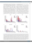
Figure 4. Transplantation of clonally cultured cells. (A) Lineage chimerism of single HSC1 cells. (B) Lineage chimerism of single-cell-derived cells in culture with stem cell factor (SCF). (C)Lineage chimerism of single-cell-derived cells in culture with SCF + granulocyte colony-stimulating factor (G-CSF). (D) Lineage chimerism of sin- gle-cell-derived cells in culture with SCF+ thrombopoietin (TPO). The percentage of the total chimerism was calculated as % (CD45.1+ cells) x 100/% (CD45.1+ cells + CD45.2+ cells). The percentage of myeloid lineage chimerism was calculated as (% CD45.1 cells) x (Mac-1/Gr-1+ cells) / (Mac-1/Gr-1+ cells + B220+ cells + CD4+ cells + CD8+ cells), in which (Mac-1/Gr-1+ cells)/(Mac-1/Gr-1+ cells + B220+ cells + CD4+ cells + CD8+ cells) was derived from CD45.1+ cells. The percentage of B- cell lineage chimerism was calculated as (% CD45.1 cells) x (B220+ cells)/(Mac-1/Gr-1+ cells + B220+ cells + CD4+ cells + CD8+ cells). The percentage of CD4 T- cell lineage chimerism was calculated as (% CD45.1 cells) x (CD4+ cells)/(Mac-1/Gr-1+ cells + B220+ cells + CD4+ cells + CD8+ cells). The percentage of CD8 T-cell lineagechimerismwascalculatedas(%CD45.1cells)x(CD8+ cells)/(Mac-1/Gr-1+ cells+B220+ cells+CD4+ cells+CD8+ cells).Micewereconsideredtoberecon- stituted with donor cells when the percentage of donor-derived cells was ≥0.2%. My-bi, Bala, and Ly-bi hematopoietic stem cells (HSC) were defined by the ratio of lymphocytes to myeloid cells (L/M ratio) in the peripheral blood 6 months after transplantation. My-bi HSC were defined by the L/M ratio <3, Ly-bi HSC were defined by the L/M ratio >10, and Bala HSC were defined by 3 < L/M < 10. Long-term (LT)-HSC were defined when the percentage of myeloid cells maintained or increased by 6 months after transplantation. Short-term (ST)-HSC were defined when the percentage of myeloid cells decreased by 6 months, with myeloid, B-lymphoid, and T-lymphoid lineage reconstitution at a time after transplantation. HPC were defined when one or two lineages lacked from the definition of ST-HSC.
haematologica | 2021; 106(6)
1653
