
Page 140 - 2021_06-Haematologica-web
P. 140
A.B. Arroyo et al.
ABCD
E
FGH
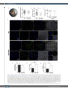
Figure 4. miR-146a deficiency accelerates carotid thrombotic occlusion. (A) Mouse model of FeCl -induced carotid arterial thrombosis. The carotid artery from wild-
3
for 2 min (WT n=24, miR-146a n=17). A Doppler ultrasound flow probe registered the blood
type (WT) and miR-146a-/- mice was isolated and exposed to 7.5% FeCl
flow continuously. (B) Basal blood flow. (C) Time to carotid thrombotic occlusion. (D) Alternatively, WT (n=7) and miR-146a-/- (n=7) mice were treated with DNase I (10 mg, i.v.) 15min before vessel injury and the carotid occlusion time was measured for a maximum of 30 min. (E) Representative images of neutrophil extracellular traps (citrullinated histone 3 [citH3]-positive nuclei) in the carotid thrombi from WT and miR-146a-/- mice analyzed by immunofluorescence. For each genotype the upper row shows the complete cross-section of the thrombosed carotid artery (bar corresponds to 100 mm) and the lower row a higher magnification of the artery wall (bar corresponds to 20 mm) and the detail of a neutrophil (bar corresponds to 5 mm) (F-H) Quantification of total infiltrated nucleated cells, citH3-positive cells and the ratio of total nucleated to citH3-positive cells in the thrombi and the adventitial layer of carotid sections from WT and miR-146a-/- mice (n=10/group). P-value calculations were performed using the unpaired Student t-test and Mann-Whitney U-test. Data represent mean ± standard error of mean, *P<0.05.
3
-/-
1642
haematologica | 2021; 106(6)
