
Page 67 - 2021_05-Haematologica-web
P. 67
Pro-leukemic effects of P2X7
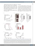
Endogenous Pbx3 in THP1 cells was suppressed by shRNA (Online Supplementary Figure S7D) with high effi- ciency (Online Supplementary Figure S7E). Inhibition of proliferation was observed in vitro, especially in THP1- hPbx3sh1 cells (Figure 7E). Furthermore, Pbx3 was also suppressed by hPbx3sh1 in leukemia cells from an AML patient with MLL translocation and high levels of P2X7 (Online Supplementary Figure S7F-H). Inhibition of prolifer- ation was also detected (Figure 7F).
These results suggest that Pbx3 mediates the pro- leukemic effects of P2X7 in murine and human models of MLL-rearranged AML.
Discussion
Nucleotide-mediated signaling is becoming a research focus in various malignancies since it has been suggested to have important roles and therapeutic potential.10 P2X7 is unique for its longest C-terminal intracellular domain of the P2X family of ATP-gated ion channels and for its formation of cytolytic pores permeable to large cations upon repeated or prolonged stimulation.27 Mutations/ polymorphisms of P2X7 have been identified in malig- nancies and abnormal expression has been detected in both malignant and microenvironmental cells.9,28
AB
C
D
E
F
Figure 7. Pbx3 mediates the effects of P2X7 on proliferation and leukemia stem cell levels in acute myeloid leukemia cells with the MLL-AF9 translocation. (A-D) Pbx3 was suppressed in MA9-P2X7 cells by shRNA against Pbx3. Equal numbers of MA9-P2X7-SC or MA9-P2X7-mPbx3sh1 cells were transplanted into recipient mice. The dynamic distribution of leukemia cells in peripheral blood (PB) is shown (A). The leukemic mice were sacrificed on day 21 and the size and weight of their spleens as well as the distribution of leukemia cells in the spleens were determined (B). Kaplan-Meier curves showing the survival of the leukemic mice (C). GFP+BFP+RFP+ leukemia cells were sorted and seeded onto 24-well plates (500 cells/well) for colony-forming assays. The proportions of the different types of colony are shown (D). (E) THP1 cells were infected with blank lentiviruses or lentiviruses carrying shRNA against Pbx3. Forty-eight hours after infection, cells were sorted, and cell proliferation was studied by an MTS assay. (F) Pbx3 was knocked down in leukemia cells from an AML patient with an MLL translocation and a high level of P2X7 expression by shRNA. Forty-eight hours after infection, cells were sorted, and cell proliferation was studied by an MTS assay. The results are from three inde- pendent experiments. Bars represent mean ± standard error of mean. *P<0.05; ***P<0.001; unpaired Student t test and one-way analysis of variance.
haematologica | 2021; 106(5)
1287
