
Page 62 - 2021_05-Haematologica-web
P. 62
W. Feng et al.
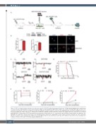
A
B
CD
E
Figure 3. P2X7 accelerates leukemia progression in the mouse model of MLL-AF9-induced acute myeloid leukemia. (A) Experimental design for the establishment of a mouse model of MLL-AF9-induced acute myeloid leukemia (AML) overexpressing P2X7 (MA9-P2X7). (B) Mice were sacrificed, and GFP+BFP+ AML cells were sort- ed by flow cytometry. The expression of P2X7 was studied by quantitative real-time polymerase chain reaction (qRT-PCR) (left), western blot (middle) and confocal microscopy (right). The relative expression of P2X7 protein normalized to GAPDH is provided. P2X7 was stained with DylightTM649 (red), and nuclei were visualized with DAPI (blue). (C) The activity of P2X7 was verified by measurement of intracellular free Ca2+ concentration upon activation. Arrows indicate the addition of 100 mΜ BzATP. (D, E) GFP+BFP+ leukemia cells (3×10⁵) were transplanted into recipient mice. Kaplan-Meier curves show the survival of leukemic mice (D), and the dis- tribution of leukemia cells in peripheral blood (PB), bone marrow (BM) and spleen was determined every 5 days (n=3) (E). Bars represent the mean ± standard error of mean. *P<0.05; **P<0.01; ***P<0.001; unpaired Student t test.
1282
haematologica | 2021; 106(5)
