
Page 208 - 2021_05-Haematologica-web
P. 208
M.V. Chan et al.
As above, data derived from the three pedigree mem- bers with the homozygous variant demonstrated a consis- tent effect upon platelet function and eicosanoid profile, irrespective of other clinical differences. Notably, the proband had an additional diagnosis of CF and administra- tion of COX inhibitors, which have anti-inflammatory effects, has been shown to inhibit the decline of lung func- tion.26 However, the proband did not show any evidence of a beneficial effect accruing from the absence of her platelet COX-1, in keeping with our understanding of the anti-inflammatory effects of NSAID being mediated pri- marily via inhibition of COX-2.27
While COX-1 protein was expressed at normal levels in platelets from the family member with the CC geno- type, it was absent in those with the GG genotype. Conversely, COX-1 was still expressed in the leukocytes irrespective of genotype. This may indicate that the PTGS1 variant is not expressed by megakaryocytes or that the variant affects the stability of the protein; i.e., that the COX-1 protein degrades more rapidly and then cannot be replenished within platelets because they lack transcriptional machinery, akin to what is observed in erythrocytes in glucose-6-phosphate dehydrogenase deficiency28. Inheritance of one copy of the mutant allele resulted in variable but never absent platelet COX-1 pro- tein levels which were sufficient to sustain function. When the variant was expressed and characterized, the recombinant protein was found to have normal enzyme activity which is consistent with the findings that in homozygous family members urinary COX-1 metabo- lites where within the normal range; i.e., implying that
despite the PTGS1 variant, COX-1 activity in tissues other than the platelet was preserved. Due to constraints in sample availability, we were unable to investigate COX-1 protein levels in other nucleated cell types in the homozygous family members.
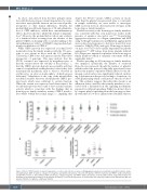
Platelet reactivity in the homozygous family members was consistent with that seen in previous studies in the presence of aspirin in vitro and in vivo.18,29,30 In particular, aggregation responses to collagen, epinephrine and ADP using both light transmission and Optimul aggregometry were reduced and responses to AA were absent but were normal to U46619 (TXA2 analogue). Homozygous knock- out mice for PTGS1 show similar impairment in platelet aggregation.31,32 ATP release from dense granules induced by collagen was impaired in platelets from homozygous family members which is similarly concordant with an aspirin-like defect29.
Platelet spreading in all homozygous family members was impaired. Specifically, the number of actin-rich filopodia was increased, though the number of platelets which reached the point of being fully spread was lower. Indeed, the number of platelets which adhered to the fib- rinogen-coated surface was significantly reduced, indicat- ing a dysfunction in the process leading to formation of a stable platelet plug which could increase the risk of bleed- ing. This evidence suggests that either this variant or an unknown defect carried by these family members is asso- ciated with a dysfunction in the signaling mechanisms required for sufficient spreading. Whilst we did not direct- ly compare platelet spreading from the homozygous fam- ily with that of low-dose aspirin-treated healthy subjects,
A
B
C
Figure 4. Effect of PTGS1 variant on platelet aggregation, secretion, and adhesion responses. (A) Aggregation responses to arachidonic acid (AA; 1 mmol/L), adeno- sine diphosphate (ADP; 10 mmol/L), collagen (0.1-3 mg/mL), epinephrine (10 mmol/L), ristocetin (1.5 mg/mL), U46619 (3 mmol/L) and TRAP-6 amide (25 mmol/L) and (B) ATP secretion to AA (1 mmol/L), ADP (10 mmol/L), collagen (3 mg/mL) and TRAP-6 amide (25 mmol/L). n=20 (healthy controls; range with median); n=1 (proband); n=2 (homozygous relatives); n=4 (unaffected relatives). (C) P-selectin expression as measured by flow cytometry in whole blood stimulated by ADP (40 mmol/L), U46619 (0.5 μmol/L) or ADP plus U46619.
1428
haematologica | 2021; 106(5)
