
Page 206 - 2021_05-Haematologica-web
P. 206
M.V. Chan et al.
reduced response to ristocetin (1.5 mg/mL; control, 69±10%; proband, 7%) which was normal in all family members tested. Other than a reduced epinephrine (10 mmol/L) response, which was found in all family mem- bers, there was no difference in other LTA responses of the unaffected family members compared to control (Figure 4A). These findings were also reflected using Optimul aggregometry where AA responses were absent, and col- lagen and epinephrine responses were severely blunted in the proband and homozygous relatives (Online Supplementary Figure 2). TRAP-6 amide (25 mmol/L)-stimu- lated ATP release was normal in all family members. AA (1 mmol/L) and collagen (3 mg/mL)-stimulated secretion, however, was below the 20th percentile in the proband and homozygous relatives (Figure 4B). Upon activation, platelets express P-selectin and undergo shape change and spreading. U46619 (0.5 mmol/L)-induced P-selectin expres- sion was similar in all individuals (Figure 4C).
The effect of the PTGS1 variant on platelet spreading The number of platelets with filopodia were increased in the proband and homozygous family members (con- trol, 9±6%; proband, 36±17%, P<0.001; homozygous rel- ative, 36±4%, P<0.01). The PTGS1 variant was also asso- ciated with a reduction in fully spread platelets (control, 45±6%; proband, 9±9%, P<0.001; homozygous relatives, 22±10%, P<0.001). Adherent platelets and number of lamellipodia were similar across all individuals tested (Figure 5). In addition, there were fewer platelets from both the proband and the homozygous relative that adhered to the fibrinogen-coated coverslips (control, 26±4%; proband, 8±2%, P<0.001; homozygous relatives,
13±3%, P<0.01).
The role of PTGS1 variant on eicosanoid production by stimulated whole blood and basal urine metabolites
Incubation of blood from healthy volunteers with colla- gen or TRAP-6 amide greatly increased the levels of TXB2 (a stable breakdown product of TXA2), 11-dehydro-TXB2 (11-dH-TXB2, a dehydrogenation product of TXB2)22, PGE2, PGD2, 15-HETE, 11-HETE and 12-HETE. In the PTGS1-deficient proband 12-HETE production was unaf- fected but there was an absence of TXB2, PGE2, PGD2 and 15-HETE (Figure 6A and B; Online Supplementary Table S1).
Despite the fact that platelets are able to synthesize
PGD , PGE and TXA from PGH , urinary metabolites for 2222
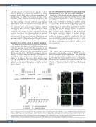
Figure 2. COX-1 protein in control, proband and relatives. (A) Western blots and quantification of cyclo-oxygenase 1 (COX-1) and glyceraldehyde 3-phosphate dehy- drogenase (GAPDH) expression in platelet lysates, isolated from the controls, the proband (IV-1), homozygous (III-1 and III-2) and unaffected (III-3, III-4, III-5 and IV- 2) family members. Representative immunohistochemical analysis of COX-1 expression in (B) control, (C) proband and (D) homozygous relative (i) platelets and (ii) leukocytes. Washed platelets were identified by tubulin (green) staining and COX-1 (magenta) was present in control but not in the proband or affected relative. In washed leukocytes, nuclear staining was confirmed by DAPI (blue), LAMP-3 (green) and COX-1 (magenta) was expressed in all samples.
these enzymes were unchanged in the proband and homozygous relatives compared to normal reference ranges. As expected, PGI2 metabolites, generated by PGI2 prostacyclin synthase from PGH in endothelial cells only
2
were all within the standard range (Figure 6C to F; Online
Supplementary Table S1). Indeed, leukocytes and endothe- lial cells are additional sources of PGD2 and PGE2 prod- ucts, respectively.
Discussion
We report autosomal recessive inheritance of a homozygous rare missense variant in PTGS1 associated with an aspirin-like platelet phenotype. This phenotype provides the opportunity to definitively assess the roles of platelet COX-1 in human platelet function, including the production of eicosanoids. This cannot be assumed from exposure of platelets from other humans to aspirin in vivo or in vitro as aspirin has effects at sites other than platelet COX-123.
A
BB
CC
DD
1426
haematologica | 2021; 106(5)
