
Page 210 - 2021_04-Haematologica-web
P. 210
J. Garcia-Reyero et al.
A
C
B
DE
FGH
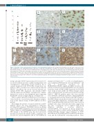
Figure 5. Phenotype of microenvironmental and neoplastic cells in plasmablastic lymphoma. (A) Scattergram illustrating the mean and range of expression values after quantification of the immunohistochemical expression of CD8, PD1 in lymphocytes and PD-L1 and CD163 in histiocyte/dendritic cell populations. (B) Representative image of a case with a mean of 36 PD-L1-positive non-neoplastic cells. (C) The same case showed a mean of 37 CD163-positive histiocytes. (D) The mean expression of CD8-positive cells in this representative case was 53. (E) PD1 identified a different T-cell subpopulation (mean of 36 PD1-positive cells in this representative example, case n. 25). (F) PD-L1 expression by neoplastic cells was identified in five out of 24 cases evaluated (20%). (G) MHCII protein/HLA (DP, DR) was, in most cases, restricted to histiocyte and endothelial cell populations. (H) MHCII protein/HLA (DP, DR) expression was identified in the neoplastic cells in three out of 25 cases tested (12%). Two of the three cases showed cytoplasmic granular and membranous staining (as illustrated in the figure) and one case had a mem- branous pattern.
notype, phospho-STAT3 expression has been found to be associated with the presence of ALK rearrangements and overexpression.20 Importantly, STAT3 activation, due to somatic mutations in the STAT3-SH2 domain may con- tribute to MYC overexpression, as previously described in DLBCL.18 In addition, one case in our series showed con- current STAT3 SH2 mutations and phospho-STAT3 (Tyr705) expression and PD-L1 overexpression, confirm- ing previous results in other lymphoma types suggesting that STAT3 activation triggers PD-L1 overexpression.21
STAT3 somatic mutations in PBL have not been previ- ously described so far and may have therapeutic implica- tions for the clinical testing of STAT3 inhibitors in these patients.
Interestingly the pattern of somatic mutations in EBV- negative disease was more heterogeneous. Mutations involving BCR activation, TLR/NFκB, histone modifying genes and the NOTCH pathway were found in eight cases (Table 1, Figure 1). MYD88pL265P mutation, involving the TIR domain of the MYD88 gene, has been previously described in activated B-cell-type DLBCL, in primary cen- tral nervous system lymphoma and in other DLBCL in
immune privileged sites22,23 as well as in lymphoplasma- cytic lymphoma/Waldenström macroglobulinemia24 and leads to downstream activation of the IRAK4/IRAK1/TRAF6 complex and NFκB activation. The pattern of mutations in CD79A/B in PBL cases was distinct from that found in DLBCL NOS. Mutations in CD79A/B were found located outside the ITAM domains related with constitutive BCR activation in activated B-cell-type DLBCL.25 NOTCH pathway genes that were mutated were NOTCH2, NOTCH1 and SGK1. NOTCH2pR2400* is a nonsense mutation that truncates the PEST domain of the NOTCH2 protein and has already been described in B- cell non-Hodgkin lymphomas, including DLBCL NOS.26 PEST domain-truncating mutations have been found in multiple tumor types and functional studies suggest that this class of mutations can be targeted with Notch inhibitors including γ secretase inhibitors.27 NOTCH1pP401L was reported in chronic lymphocytic leukemia in a previous study28 and lies within the calcium- binding EGF-like domains repeat. Mutations in SGK1 involved the SGK1pS451F and SGK1pA380V point muta- tions and the SGK1pK136* truncating mutation. These
1126
haematologica | 2021; 106(4)
