
Page 209 - 2021_04-Haematologica-web
P. 209
Oncogenic somatic mutations in plasmablastic lymphoma.
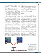
tion of PBL cases has STAT3 activation due to somatic mutations in the STAT3-SH2 domain that may increase MYC expression, as previously described in DLBCL18 (Figure 4).
Phenotype of the immune microenvironment and neoplastic cells in plasmablastic lymphoma
We quantified the expression of CD163 and PD-L1 in histiocytic/dendritic cells in the cases of PBL. The mean expression of PDL1 was 33 nuclei per HPF (range, 1.67-61) and the mean expression of CD163 was 38 nuclei per HPF (range, 2-84) (Figure 5). The correlation between CD163 and PD-L1 expression was statistically significant (Pearson 0.6, P<0.05), suggesting that PD-L1-positive cells are histiocytes in PBL. There was not a significant difference in the content or distribution of CD163 and PD-L1-posi- tive histiocytes between EBV-positive and EBV-negative cases (Mann-Whitney test, P>0.05).
CD8-positive and PD1-positive T-cell subpopulations were quantified. The mean number of CD8-positive lym- phocytes was 52 nuclei per HPF (range, 1-117) and the mean number of PD1-positive lymphocytes was 32 nuclei per HFP (range, 0-76). There was a significant difference in the distribution of CD8 and PD1-positive cell subsets (Wilcoxon test, P<0.001) consistent with different cell populations. The Pearson correlation value was however statistically significant (Pearson 0.59, P<0.05). There was no significant difference in the content and distribution of CD8 or PD1-positive lymphocytes between EBV-positive and EBV-negative cases (Mann-Whitney test, P>0.05) (Figure 5).
PD-L1 was expressed by tumor cells in five out of 24 (20%) cases evaluated (mean 59 nuclei per HPF; range, 25- 98). Four out of five PD-L1-positive cases (in the neoplastic cells) were EBV-positive. Fourteen EBV-positive PBL cases were negative for PD-L1 in the tumor cells. Thus four out of 18 (22%) EBV-positive PBL cases were PD-L1-positive, while one out of six (16%) EBV-negative cases was PD-L1- positive. Thus, there was no association between EBV infection by tumor cells and PD-L1 expression, since most of the EBV-positive cases were PD-L1-negative (P=non- significant) (Figure 5). Interestingly one case with STAT3 SH2 mutations showed concurrent PD-L1 and phospho- STAT3 (Tyr705) expression. PD-L1 expression data were
not available for the other STAT3 SH2-mutated cases to test this association.
Consistent with previously published data,9 MHCII pro- tein/HLA (DP, DR) was virtually absent in PBL. Only three cases out of 25 tested were positive (12%, mean 349 nuclei per HPF; range, 284-440). Two cases showed a membranous and cytoplasmic granular pattern and the other a membranous pattern. All three cases were EBV- positive. The other 22 cases were completely negative for HLA expression in tumor cells (Figure 5).
Discussion
In this study we characterized the genetic profile of a series of cases of PBL using targeted exonic NGS, any cor- relations with EBV infection and the expression of immune checkpoint proteins in both the neoplastic popu- lation and tumor microenvironment. We found that genet- ic abnormalities (including translocations, amplifications and point mutations) in the MYC gene were the most common genetic event in PBL. In addition to previously described translocations, involving IGH and MYC,10,11 here we found that a few cases may have MYC amplification, confirming our previous observations.12 Both MYC translocations and amplifications lead to a significantly increased expression of MYC protein. Interestingly we also identified MYC point mutations, mainly consisting of transversions and transitions at C:G pairs and involving exon 2 and, in the case of MYCp79S mutation, the WRCY consensus motif. All these features are consistent with a mechanism related to aberrant somatic hypermutation.16 The oncogenic effect of these point mutations does, how- ever, remain unclear.
We also found that 16% of our cases (5 cases) carried recurrent somatic mutations in the oncogene STAT3, pref- erentially involving the SH2 domain of the protein. Interestingly these mutations were restricted to EBV-posi- tive PBL. Here we demonstrate that these mutations led to increased expression of phospho-STAT3 (Tyr705).
STAT3 mutations and phospho-STAT3 overexpression have been found very rarely in DLBCL NOS (6% accord- ing to Ohgami et al.19). In cases of ALK-positive large B-cell lymphomas, which commonly show a plasmablastic phe-
Figure 4. MYC protein overexpression in plasmablastic lymphoma. MYC protein over- expression is due to rearrangements involv- ing MYC in a significant proportion of cases of plasmablastic lymphoma (69% in these series). Most translocations fuse MYC to IGH and a few cases may show amplifications of the MYC gene. In addition, we found that some cases of plasmablastic lymphoma have STAT3 activation due to somatic muta- tions in the STAT3-SH2 domain which may increase MYC expression.
haematologica | 2021; 106(4)
1125
