
Page 208 - 2021_04-Haematologica-web
P. 208
J. Garcia-Reyero et al.
ifying genes and the NOTCH pathway were found in eight cases (Table 1, Figure 1). These mutations involved CD79pAW76*, CD79BpD34N, MYD88pL265P, NOTCH1pP401L, NOTCH2pR2400*, SGK1Kp136*and EP300pM2010I/EP300pR1731H. The NOTCH pathway was affected by somatic mutations in NOTCH2 (1 case), NOTCH1 (1 case) and SGK1 (2 cases). Other mutations found were SMARCA4pR1005Q and TP53pR273H. Of note, two cases, both EBV-negative, had mutations in the BRAF gene, one case with the canonical activating BRAFpV600E mutation and the other with a BRAFpG469A mutation in the ATP binding site.
STAT3 mutations are associated with constitutive phospho-STAT3 (Tyr705) activation and MYC protein overexpression is related to MYC rearrangement status
Expression of phospho-STAT3 (Tyr705) protein was quantified immunohistochemically in 20 cases with avail- able mutational data. Mean phospho-STAT3 expression was 48 nuclei per high power field (HPF; 40x) in these 20 cases. Mean expression for two out of four SH2 domain- mutated cases with available immunohistochemical data was 249 nuclei per HPF. Mean phosho-STAT3 expression for STAT3 wild-type cases was 28 nuclei per HPF. Mean phospho-STAT3 expression for the single non-SH2 STAT3- mutated sample was 40 nuclei per HPF. Thus, STAT3 SH2 domain mutations (STAT3pY640F, STAT3pM648L, STAT3pG618R, STAT3pN647I) were associated with over- expression of phospho-STAT3, as determined by immuno- histochemistry of tissue samples (Figure 2B).
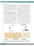
MYC protein was consistently expressed in all the cases (range, 59-236 nuclei per HPF; mean 236), irrespective of the presence of MYC translocations, as previously report-
ed.12,17 However, significant differences in the level of MYC expression were found, according to MYC gene sta- tus. MYC-translocated (14 cases) and -amplified cases (2 cases) had, as expected, higher MYC protein expression than cases without MYC rearrangements (7 cases). The mean number of positive nuclei per HPF was 109 in non- rearranged cases versus 282 in MYC-rearranged cases (Mann-Whitney test, P<0.0001) (Figure 3).
Mean MYC protein expression in 22 cases with avail- able data was 236 nuclei per HPF, which was significantly higher than the mean 48 nuclei per HPF in the cases of phospho-STAT3 protein expression (Wilcoxon test, P<0.001). There was no correlation between the levels of expression of the two proteins (Pearson test, non-signifi- cant). Due to the high prevalence of MYC translocations and amplification in PBL and the relatively low levels of phospho-STAT3 expression and absence of correlation between the proteins, it is unlikely that STAT3 activation contributed to MYC overexpression in most cases. However, one of our cases with STAT3 SH2 domain muta- tions and absence of MYC translocation by FISH showed high levels of both phospho-STAT3 and MYC proteins, without detectable PRDM1/Blimp1 mutations, suggesting that MYC overexpression might be related with STAT3 activation by mutations in rare cases of PBL.
In summary, MYC protein overexpression is due to rearrangements involving MYC in a significant proportion of cases of PBL (69% in our series). Most translocations fuse MYC to IGH and a few cases may show amplifica- tions of the MYC gene. Both alterations lead to MYC pro- tein overexpression. Genetic alterations in the MYC regu- latory domains of PRDM1/Blimp1 may also contribute to its overexpression.12 In addition here we show that a frac-
AB
C
Figure 3. MYC protein expression in plasmablastic lymphoma. (A) MYC protein was consistently expressed in the cases of plasmablastic lymphoma. (A) Mean MYC protein expression in 22 cases with available data was 236 nuclei per high power field (HPF), which was significantly higher than the mean of 48 nuclei per HPF in the case of phospho-STAT3 protein expression (Wilcoxon test, P<0.001). MYC translocated cases (n=14) and MYC amplified cases (n=2) had higher MYC protein expression than cases without MYC rearrangements (n=7) (Mann-Whitney test, P<0.0001). (C) Representative microphotographs of MYC protein expression in plas- mablastic lymphoma.
1124
haematologica | 2021; 106(4)
