
Page 196 - 2021_04-Haematologica-web
P. 196
G. Juban et al.
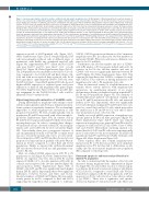
Figure 3. (previous page) Gata1s cells fail to produce erythroid cells and mature megakaryocytes. (A) Micrographs of May-Grunwald-Giemsa-stained cytospins of fluorescence-activated cell sorting (FACS)-purified double negative (DN) (kit–CD41–) cells at day 10 (d10). Genotype of cells is indicated above. Scale bars represent 25 mm. (B) Bar plot of number of erythroid cells (Ter119+) and myeloid cells (Gr1+ and/or Mac1+) within the DN population at d10 of culture in BirA, bioG1 and bioG1s cultures. 250,000 total cells analysed by FACS in each case. (C) Heatmap of mRNA expression of selected erythroid (top), myeloid (middle) and megakaryocytic (bot- tom) genes (in rows) in DN BirA (left), bioG1 (middle) and bioG1 (right) cells at d10. Data from two independent biological replicates is shown. (D) Representative histogram (three independent experiments were performed) showing size (forward scatter [FSC-A], top panel) and granularity (side scatter [SSC-A], bottom panel) assessed by flow cytometry. Data from BirA (left), bioG1 (middle), bioG1s (right) from P2, DN, P3, P4 populations is shown. In P4, numbers indicate the mean per- centage ±1 standard deviation (SD) of cells within the gate. (E) Bar plot showing the number of cells expressing CD42b at d12 from DN, P1, P2, P3 and P4 popula- tions from genotypes indicated (three independent experiments). (F) Top, representative micrographs of acetylcholinesterase (AChE) staining of FACS-purified P1 to P4 d10 BirA, bioG1 and bioG1s populations. Scale bars represent 10 mm. Bottom, quantitation of AChE staining, from 500 cells, analyzed from three independent experiments. Bean-plot of staining intensity expressed in arbitrary units (AU) (y-axis) for each population (P1 to P4) from BirA, bioG1 and bioG1s cells. (G) Heatmap showing the fold change in expression of selected megakaryocytic genes (indicated on the right) in FACS-purified P1, P2, P3 and P4 at d10 (columns). Data from two independent biological replicates is shown. Genotype of the cells is indicated below the heatmap. (H) Hierarchical clustering using mRNA data from (G). *P<0.05, **P<0.01 and ***P<0.001 between bioG1s and BirA. #P<0.05, ##P<0.01 and ###P<0.001 between bioG1s and bioG1. §P<0.05 and §§P<0.01 between P4 and P2. $$P<0.01 and $$$P<0.001 between P4 and P3.
expression profile of FACS-purified cells (Figure 3A-C; Online Supplementary Figure S4A-F). Morphologically, DN cells were primarily erythroid cells, at different stages of maturation, with hardly any granulated myeloid cells (Figure 3A). Approximately 50% of BirA and bioG1 DN cells were Ter119+ and 5% were Mac1+, Gr1+ or both Mac1+Gr1+ (Figure 3B; Online Supplementary Figure S4A-C). The DN population was markedly reduced in bioG1s cul- tures compared to bioG1 (24-fold) and BirA cultures (16- fold) and with more myeloid than erythroid cells. In all three genotypes approximately 50-60% DN cells were Ter119–Gr1–Mac1–. Given FACS-purified DN cells showed higher mRNA expression of erythroid genes and lower expression of myeloid and megakaryocytic genes (Figure 3C; Online Supplementary Figure S3D-F), one possible line- age assignment for the Ter119–Gr1–Mac1– cells could be immature Ter119– erythroid cells.
Altered megakaryocytic maturation of bioGATA1s cells
During differentiation, megakaryocytes enlarge consid- erably, acquire granules and develop a demarcation mem- brane system for proplatelet formation. We used multiple approaches to study megakaryocyte maturation as cells progressed from P2 to P4. First, morphologically, cells in populations P1 and P2 were small, with a blast morpholo- gy (Online Supplementary Figure S5A). In contrast, cells in P3 and P4 were larger, particularly P4 which were matur- ing megakaryocytes. In order to quantify these changes, we measured cell size (forward scatter[ FSC-A]) and gran- ularity (side scatter [SSC-A]) by flow cytometry (Figure 3D). Concordantly, there was a progressive increase of size and granularity from P2 to P3 and P4. A similar trend was also seen in the mutant cells, but to a lower extent. Closer inspection of FSC and SSC profiles showed a lower proportion of larger and more granular cells in the P4 pop- ulation in bioG1s compared to control BirA and bioG1 populations. Finally, as expected the erythroid-dominant DN cells showed decreased cell size and granularity com- pared to P2 cells.
Next, we studied CD42b expression in DN, P1 to P4 populations at d12 (Figure 3E; Online Supplementary Figure S5B-E). As expected, very few cells in DN, P1 and P2 expressed CD42b (<4%; absolute numbers <200 cells). In contrast, and as expected, the absolute number (Figure 3E) and proportion (Online Supplementary Figure S5E) of CD42b+ cells in P3 and P4 were significantly much higher than in DN, P1 and P2. Importantly, there were significant differences between bioG1s and BirA and bioG1. Absolute numbers of CD42b+ in P3 (Figure 3E) were signif- icantly greater in bioG1s compared to BirA and bioG1 supporting the hypothesis that compared to wild-type
GATA1, GATA1s promotes proliferation of kitlo immature megakaryocytes (P3). In contrast, the absolute numbers of mature kit–CD42b+ P4 bioG1s cells were no different com- pared to bioG1 and BirA.
Furthermore, the absolute number and ratio of CD42b+ cells in P4 relative to P3 was greater in BirA and bioG1. In contrast, in bioG1s the absolute number and ratio of CD42b+ cells was not significantly different between P3 and P4 (Figure 3E; Online Supplementary Figure S5E). This supports the hypothesis that GATA1s, compared to wild- type GATA1, is less effective at driving maturation of P3 megakaryocytic cells to P4 megakaryocytic cells.
Next, we measured activity of acetylcholinesterase, an enzyme whose activity increases with megakaryocyte maturation, by quantitating intensity of an acetyl- cholinesterase driven cytochemical reaction in purified P1, P2, P3 and P4 populations (Figure 3F). The intensity of acetylcholinesterase-induced cytochemical staining was low in P1 and P2 and increased in P3 cells, and increased further, in P4 cells. Importantly, there was significantly lower cytochemical staining in P3 and P4 in bioG1s com- pared to control BirA and bioG1 cells, which may reflect the smaller size of P3 and P4 bioG1s cells and/or differ- ence in maturation state of bioG1s cells.
Finally, we tested mRNA expression of megakaryocyte specific genes in P1-P4 in all three genotypes (Figure 3G; Online Supplementary Figure S4E-F). There was reduced expression of megakaryocyte genes in P3 and P4 in bioG1s compared to BirA and bioG1 cells (Tubb1, Factor V, Pbbp, Gp9 and Hsd3b6). Hierarchical clustering analysis con- firmed that bioG1s P3 and P4 cells were transcriptionally more closely related to the more immature P1 and P2 cell populations than P3 and P4 from GATA1 wild-type cells (Figure 3H; Online Supplemental Figure S5F).
Taken together, these data confirm megakaryocytes mature from P1 to P4. BioG1s produce more immature megakaryocytes (P3) but they fail to differentiate as effi- ciently into the most mature megakaryocyte population (P4) compared to wild-type cells.
Decreased apoptosis and increased proliferation in mutant P3 population
In order to understand why the number of bioG1s cells increased during megakaryocytic differentiation (Figure 1G; Figure 3E), we asked if this was due to reduced apop- tosis (Figure 4A-B) and/or increased cycling of cells (Figure 4C-D) in bioG1s compared to control BirA cells. We per- formed flow analyses in P1-P4 sub-populations at d8 (for apoptosis) or d9 (for cell cycle) (2 or 3 days after re-culture of kithiCD41lo). There was a significant, and specific, decrease in Annexin V+ cells in bioG1s P3 cells. There was
1112
haematologica | 2021; 106(4)
