
Page 145 - 2021_04-Haematologica-web
P. 145
DEX and DAS impair in vivo T-ALL propagation
ABC
DE
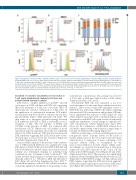
Figure 4. Lymphocyte cell-specific kinase (LCK) knockdown leads to cell cycle arrest in T-cell acute lymphoblastic leukemia (T-ALL) cell lines and patient-derived xenograft (PDX) cells. (A-C) Cell cycle status was determined by flow cytometry using Hoechst 33342 in cell lines Jurkat (A and C), MOLT4 (B and C), and SUPT1 (C) 7 days after transduction with shLCK#3 or shNTC expression vectors. (D) PDX L963 cells were lentivirally transduced with shLCK#3 or shNTC expression constructs. Phosflow analysis of total LCK and p-Y416SRC was performed 8 days later. (E) shLCK#3 and shNTC transduced PDX L963 cells were loaded with CTV and cultured for 13 days. Flow cytometric analysis of CTV incorporation (cell divisions) was performed and demonstrated progressive reduction in cell number after 4-6 cell divisions after LCK knockdown relative to control knockdown. Student's t-test: *P<0.05, **P<0.01, ***P<0.005.
Dasatinib re-sensitizes dexamethasone resistance in T-cell acute lymphoblastic leukemia cell lines and patient-derived xenograft samples
DAS leads to complete inhibition of p-Y416SRC and cell cycle arrest in T-ALL cell lines and PDX cells, suggesting that DAS treatment of T-ALL has a cytostatic effect. In clinical practice, effective eradication of T-ALL relies on the application of combinatorial treatment. LCK inhibi- tion has previously been shown to sensitize chronic lym- phoid leukemia (CLL) to DEX and induce cell death.23 We thus went on to investigate potential synergy between LCK inhibition and DEX, as DEX is universally used for treatment of ALL. The cell viability of SUPT1 and CUTLL1, in the presence of DEX, was evaluated after knockdown of LCK. Whereas the cell viability of mock transduced and non-targeting control cells was minimally affected by DEX treatment, LCK knockdown increased DEX sensitivity suggesting that LCK protein and/or activ- ity levels play a crucial role in GC resistance (Figure 6A and Online Supplementary Figure S6A).
A more detailed analysis of the potency of the combina- tion of DEX and LCK inhibition was examined by using DAS instead of the LCK knockdown. DEX (0-600 nM) and DAS (0-50 mM) were titrated along a dose matrix and cell viability was determined. Synergy for individual drug combinations was determined using Combenefit.22 The matrix revealed drug synergy at concentrations which are clinically achieved, i.e., 100 nM for DEX and 264 nM for DAS (Figure 6B and Online Supplementary Figure S6B).24,25 Bioinformatic analysis of all ten T-ALL cell lines revealed a statistically significant enrichment of drug synergy at clin-
ically relevant concentrations. This synergy was observed at 8-110 nM of DEX and 0.223-4.5 μM of DAS (Online Supplementary Figure S6C and D).
Subsequently, PDX cells were expanded ex vivo for 1 week and exposed to the same drug combinations in dose matrices. These assays verified the synergistic action of DEX+DAS in a wide range of PDX cells, whilst confirming that increased DAS concentrations and resultant LCK inhi- bition augmented the response to DEX (Figure 6C and Online Supplementary Figure S6E). Combined analysis of all drug matrices with PDX cells again revealed a statistically significant enrichment of drug synergy at clinically rele- vant concentrations (Online Supplementary Figure S6F). Moreover, the combination of DEX+DAS induced more cell death compared with control vehicle or single drugs as revealed by Annexin V/PI staining (Figure 6C).
DEX has a wide range of actions, including genomic and non-genomic effects. Genomic effects are the result of nuclear translocation of the GC receptor and subsequent transactivation or repression of genes containing a GC response element (GRE), as exemplified by the Glucocorticoid-Induced Leucine Zipper (GILZ) gene. Accordingly, we observed strong induction of GILZ gene expression after DEX exposure in the T-ALL cell line Jurkat and five PDX samples tested (Figure 6D and Online Supplementary Figure S6G). This response was significantly enhanced when combining DEX with knockdown of LCK (Figure 6D) or DEX+DAS in a range of T-ALL cell lines and PDX samples, suggesting that LCK inactivation augments DEX-induced gene transcription and reverses DEX resist- ance (Figure 6D and Online Supplementary Figure S6G).
haematologica | 2021; 106(4)
1061
