
Page 143 - 2021_04-Haematologica-web
P. 143
DEX and DAS impair in vivo T-ALL propagation
A
BC
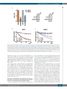
Figure 2. Knockdown of lymphocyte cell-specific kinase (LCK) reduces propagation of T-cell acute lymphoblastic leukemia (T-ALL) cell lines in vitro. SUPT1 (A and B) and MOLT4 (A and C) cells were lentivirally transduced with shNTC (non-targeting control), shLCK#1, shLCK#2, shLCK#3, shPTCRA#1, or shZAP70#1 expression constructs. (A) Knockdown efficiency of LCK at mRNA level (left) and protein level (right) after 6 days. Whole cell lysates were probed for total LCK and GAPDH in western blot analysis. (B and C) T-ALL cell lines SUPT1 (B) and MOLT4 (C) transduced with GFP-expressing shLCK (blue), shZAP70 (purple), shPTCRA (red) or shNTC (black) constructs were seeded in a 1:1 ratio with non-transduced parental cells in vitro. Cells were cultured and analyzed repetitively by flow cytometry for the pres- ence of GFP+ cells over a time period of 30 and 40 days for SUPT1 and MOLT4, respectively. A relative GFP expression of 1 denotes a mixture of 50% GFP+ cells with 50% parental cells (ratio 1:1). A value of 0.5 refers to 25% of GFP+ cells and 75% parental cells (ratio 1:4). Student's t-test: ****P<0.001.
Supplementary Figure S3A). Genomic DNA (gDNA) was extracted from L963 cells isolated from bone marrow and spleen after mice became symptomatic (week 11). ShRNA sequencing indicated that shLCK#3 represented the most significantly depleted shRNA construct in vivo (Figure 3A, Online Supplementary Table S3 and Online Supplementary Figure S3B).
To assess the effect of LCK knockdown on engraftment fitness, MOLT4 cells were transduced with lentiviral vec- tors encoding either red fluorescent protein RFP/shNTC (non-targeting control) or GFP/shLCK#3. Equal propor- tions of cell populations were transplanted into NSG mice (n=5). Leukemia cells were isolated from spleen, bone marrow and liver once mice were symptomatic (day 26). Flow cytometric analysis of the leukemic cell population established that cells carrying shNTC had a clear compet- itive engraftment advantage over cells with LCK knock- down in all mice tissues sampled (Figure 3B and Online Supplementary Figure S3C).
Knockdown of lymphocyte cell-specific kinase leads to cell cycle arrest in T-cell acute lymphoblastic leukemia cell lines and patient-derived xenograft samples
Next we investigated the mechanisms underlying the
defect in proliferation, survival and engraftment observed after LCK knockdown. Jurkat, MOLT4 and SUPT1 cells were transduced with shLCK#1/#3 and cell cycle analyses performed. In all cell lines, we observed significant cell cycle arrest with an increase in G0/G1 phase and decrease in S phase after LCK knockdown (Figure 4A-C and Online Supplementary Figure S4A).
ShLCK#3 led to decreased protein levels of total LCK and activated p-Y416SRC in cell lines, suggesting activation status of LCK is associated with cell cycle arrest. In PDX L963, LCK knockdown led to a 45% reduction in total LCK expression, as well as a 71% reduction in p-Y416SRC as assessed by Phosflow (Figure 4D). This knockdown resulted in a decrease in S phase over time compared to control (Online Supplementary Figure S4B). The proliferative behavior of PDX cells was analyzed after labeling with cell trace violet (CTV). PDX L963 cells were transduced with shRNA constructs targeting LCK or a non-targeting con- trol (NTC) and co-cultured with OP9-DL1 feeder cells for 13 days. The LCK knockdown cells showed restricted pro- liferation compared to the control cells (Figure 4E). Confirmatory siRNA knockdown of LCK was undertaken in PDX samples LK203 and L963. Knockdown of total and activated LCK was confirmed by Phosflow. Cell cycle
haematologica | 2021; 106(4)
1059
