
Page 188 - 2021_03-Haematologica-web
P. 188
J. Chen et al.
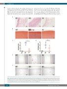
features: chronic synovitis and cartilage destruction. An enlarged synovial membrane accompanied by inflamma- tory infiltrations was observed in 75% (3 of 4) of the pF9 KO pigs, and a thin synovial membrane with subsynovial adipose tissue was seen in 75% (3 of 4) of the hF9 KI pigs (Figure 4A). In addition, severe cartilage destruction was
A
B
observed in 75% (3 of 4) of the pF9 KO pigs, while mild chondrocyte and proteoglycan losses were also found in 75% (3 of 4) of the hF9 KI pigs (Figure 4B). The arthropa- thy score was applied to the histological analysis. There was a significant difference in synovial change between pF9 KO pigs and hF9 KI pigs and almost no signs of syn-
CD
E
F
Figure 4. Histological analysis of the ankle joints of pF9 knockout (KO) pigs and hF9 knockin (KI) pigs. (A) An enlarged and inflamed synovial membrane was shown in the pF9 KO group, while the thin synovial membrane with subsynovial adipose tissue in the hF9 KI group was comparable to that in the WT group. (B) Safranin O staining was performed to show proteoglycan loss and chondrocyte apoptosis in pF9 KO pigs and hF9 KI pigs. (C) Histological sections were scored to assess synovitis (0-3). (D) Histological sections were scored to assess cartilage degradation (0-3). (E) pF9 KO pigs exhibited coagulated blood in the ankle joint cavity. (F) Joint surface of the ankle joints. WT, n=4; KO, n=4; KI, n=4. Scale bars: 50 mm. ***P<0.0001, ns: not significant.
834
haematologica | 2021; 106(3)
