
Page 186 - 2021_03-Haematologica-web
P. 186
J. Chen et al.
ABC
DEFG
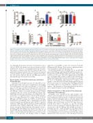
Figure 2. The analysis of blood coagulation and factor IX (FIX) levels. (A) FIX activities in the plasma of pigs and normal humans. The activity was calculated on the basis of a standard curve generated with normal human plasma and is expressed as a percentage of FIX activity in normal human plasma (FIX activity: WT: approx. 429%; KO: approx. 23.8%; KI: approx. 58%). (B) Measurement of the APTT. (C) Measurement of the PT. (A-C: Human, n=3; WT, n=4; KO, n=4; KI, n=3). (D) quantita- tive-real-time-polymerase chain reaction (qRT-PCR) detection of mRNA expression levels of porcine factor IX in the livers from pF9 KO pigs and hF9 KI pigs. (E) qRT- PCR detection of mRNA expression levels of human factor IX in the livers from pF9 KO pigs and hF9 KI pigs. (F) Western blotting of liver tissues with an antibody against human FIX and porcine FIX. GAPDH served as the control. Quantitation of the ratios of FIX to GAPDH is shown. Western blot analysis was repeated independ- ently at least three times. (G) Determination of the FIX concentration in the plasma of pF9 KI pigs using human plasma as a control. The data are presented as the mean±standard deviation and were analyzed using an unpaired t-test with Welch's correction using GraphPad Prism software. The error bars represent the standard deviation. **P<0.005, ***P<0.0001, ns: not significant (vs. the WT pigs). #P<0.05, ##P<0.005, ###P<0.0001.
S3). Similarly, the positive cell clones were mixed as donor cells for SCNT; the detailed records are shown in Online Supplementary Table S2. Genotyping by PCR and sequenc- ing analysis confirmed that the five delivered pigs were genetically identical and the hF9 CDS had been inserted correctly (Figure 1E and F and Online Supplementary Figure S4). The results indicated that the pF9 KO pigs and hF9 knockin (KI) pigs developed successfully.
Blood analysis of the pF9 knockout pigs and human F9 knockin pigs
Clinically, as an X-linked recessive disorder, HB is com- mon in men. Thus, we focused on male pigs to investigate their pathological and behavioral changes. Four pF9 KO pigs, four WT pigs, and three hF9 KI pigs were included in the blood analysis. FIX activity was severely decreased in pF9 KO pigs, while it was partly increased in hF9 KI pigs compared with the activity in pF9 KO pigs (Figure 2A). When FIX activity in porcine plasma was expressed as the percentage of coagulation factor activity in WT pig plas- ma, it was approximately 5.5% (range: 4.5-6.2%) in pF9 KO pigs and 13.5% (range: 12.3-15.0%) in hF9 KI pigs. Furthermore, pF9 KO pigs had a significantly prolonged activated partial thromboplastin time (APTT) compared with that of WT pigs, and the insertion of hF9 CDS partly ameliorated the condition (Figure 2B). There was no dif- ference among the different groups with regard to the pro- thrombin time (PT) (Figure 2C). Apart from this, two of the pF9 KO pigs had significantly decreased red blood cell (RBC) counts and hemoglobin (Hb) levels, as well as lower levels of total proteins and albumin (Table 1). Higher
white blood cell (WBC) counts were found in both pF9 KO pigs and hF9 KI pigs. There were no significant differ- ences in platelet counts among all groups.
Porcine FIX mRNA was extremely low in all pF9 KO pigs and hF9 KI pigs, and human FIX mRNA was effective- ly transcribed in hF9 KI pigs (Figure 2D and E). FIX protein synthesis in livers also decreased significantly in pF9 KO pigs, and human FIX could be synthesized in hF9 KI pigs (Figure 2F). Human FIX could be successfully secreted into the blood of hF9 KI pigs, approximately 9% (approx. 90.4 ng/mL) of the FIX level in normal human plasma (Figure 2G). Based on the above analysis, the pF9 KO pigs showed lower FIX expression and exhibited coagulation abnor- malities. The insertion of hF9 partly alleviated the abnor- mal coagulation function in the pF9 KO pigs.
Clinical observations of the porcine F9 knockout pigs and human F9 knockin pigs
All piglets were separated from their mothers after delivery, and each piglet was housed individually by arti- ficial suckling in a cage the insides of which had cushioned buffers to protect the piglets and avoid trauma (Figure 3A). Clinical observations of ten cloned pigs, including five pF9 KO pigs, three hF9 KI pigs, and two WT pigs were per- formed from birth to 8 weeks of age. No bleeding episodes were recorded in WT pigs throughout the obser- vation period. All pF9 KO pigs had multiple spontaneous bleeding episodes, while spontaneous bleeding with a lower frequency occurred in 67% of hF9 KI pigs (2 of 3); no bleeding episode was found in one hF9 KI pig (Online Supplementary Table S3). Most of the bleeding occurred in
832
haematologica | 2021; 106(3)
