
Page 185 - 2021_03-Haematologica-web
P. 185
Porcine model of hemophilia B
reconstructed embryos were transferred into nine surro- gate pigs (Online Supplementary Table S2). Ultrasonography was performed 30 days post transfer and revealed that four recipients had become pregnant. All pregnancies were maintained to term, and 12 founder (F0) pigs (8 males; 4 females) were delivered. Genotyping by PCR and DNA sequencing identified that eight male pigs carried an F9 deletion and four female pigs had a het- erozygous genotype (Figure 1B and C). The female F0 pigs were used to mate with the male F0 pigs to generate F1 pigs. The breeding gave rise to seven F1 pigs, including two male WT pigs, two male F9-deletion pigs, and three
female heterozygous pigs (Online Supplementary Figure S2). Meanwhile, we wondered whether the insertion of a single copy of the human F9 (hF9) gene could alleviate the phenotype in porcine F9 (pF9) knockout (KO) pigs. Thus, in addition to the previously identified two sgRNAs, a donor vector that carries the hF9 CDS flanked by 1-kb homology sequences on both sides was also generated (Figure 1D). By co-transfecting the Cas9/sgRNA plasmids and the donor vector into male fetal pig fibroblasts, we obtained five positive cell clones, and the subsequent sequencing results showed that the hF9 CDS was inserted correctly into the pF9 locus (Online Supplementary Figure
AB
C
DE
F
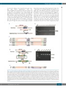
Figure 1. Generation of pF9 knockout (KO) pigs and hF9 knockin (KI) pigs. (A) Schematic diagram of the strategy used to generate the F9 KO pigs. Two sgRNAs were used to target the 5’ untranslated region (UTR) and exon 1 (E1) of pF9 to promote DNA breaks. (A) The primer pair F1/R1 was used to identify the knockout of pF9. (B) The targeting regions were amplified using genomic DNA derived from the piglets. The gene is located on the sex chromosome: eight male piglets (F0-1, F0-2, F0-3, F0-6, F0-7, F0-8, F0-9, F0-10) presented single deletion bands, whereas the other four female piglets (F0-11, F0-12, F0-13, F0-14) who have two F9 alleles presented heterozygous bands. (C) The exact genotypes of pF9 KO pigs were determined by Sanger sequencing. The targeting sequences of the sgRNAs are marked in red, while the protospacer adjacent motifs (PAM) are marked in green. The mutated sequences of male pigs are presented above the line, and the corresponding sequences of female pigs are arranged below. KO-T1: the genotype of a 117 bp deletion; KO-T2: the genotype of a 118 bp deletion. (D) Strategy used for the CRISPR/Cas9-mediated knockin of hF9 into the endogenous pF9 locus via homologous recombination. The donor DNA consisted of hF9 coding sequences (CDS) and homologous porcine sequences (left arm and right arm), which were used to integrate hF9 into the porcine genome. 5’ HA: 5’ homologous arm (1.0 kb); 3’ HA: 3’ homologous arm (1.0 kb). (E) The newborn hF9 KI piglets were identified via polymerase chain reaction using the primer pair F1/R1. (F) The genome sequences of hF9 KI pigs indicate the successful insertion of the hF9 CDS. The modified bases that avoid recutting after homologous recombination are underlined and marked in purple.
haematologica | 2021; 106(3)
831
