
Page 145 - 2021_03-Haematologica-web
P. 145
Iron deficiency and thrombosis
A
BCD
E
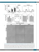
Figure 7. Iron deficiency increases the expression of P-selectin, without altering platelet aggregability or static adhesion. (A) Mean fluorescence intensity of P-selec- tion expression on platelets before and after stimulation with thrombin (0.0156 U/mL, 0.25 U/mL). Error bars: mean ± standard deviation (SD) (n=4 per group). Representative histograms show the population shift. (B) Plasma P-selectin concentration determined by enzyme-linked immunosorbent assay (n=4 per group). (C) Platelet Function Analyzer closure time, a measurement of platelet adhesion under shear flow (n=5 per group), depicted as seconds until occlusion, or no occlusion. (D) Area under the curve measured from Multiplate whole blood impedance aggregometry after stimulation with ADP or collagen (n=4-5 per group). (E) Representative images of washed platelets adhering to a fibrinogen- or collagen-coated surface. *P<0.05. Error bars: mean ± SD. P-sel: P-selectin; MFI: mean fluo- rescent intensity; sP-sel: soluble P-selectin; PFA-100 CT: Platelet Function Analyzer 100 closure time; AUC: area under the curve; Con: animals fed a normal diet and given placebo injections; Def: animals fed an iron-deficient diet; Def+Fe: animals fed an iron-deficient diet and then given injections of ferric carboxymaltose.
haematologica | 2021; 106(3)
791
