
Page 140 - 2021_03-Haematologica-web
P. 140
K. Jimenez et al.
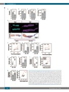
ABCD
E
FGHI
JKLM
NO
Figure 2. Iron deficiency augments venous thrombotic tendency. In experiment 1, venous thrombo- sis was induced by stenosis of the inferior vena cava after animals had been on an iron-deficient diet (Def) for 7 weeks. A control group (Con) was fed a normal diet. (A-D) Hematologic parameters meas- ured prior to surgery (n=7 per group); hemoglobin (A), mean corpuscular volume (B), hematocrit (C) and platelet count (D). (E) Representative three-dimensional images reconstructed from high fre- quency ultrasound scans at 3 and 4 h after ligation; and corresponding Carstairs staining of thrombi collected post-mortem. (F, G) Thrombus volume at 3 and 4 h after ligation (F) and the change in vol- ume over time (G) (n=5 per group, error bars: mean ± standard deviation [SD]). (H) Final thrombus volume at 4 h (n=6-7 per group). (I, J) Thrombus length at 3 and 4 h after ligation (I) and the change in length over time (J) (n=5 per group, error bars: mean ± SD). (K) Final thrombus length at 4 h (n=6- 7 per group). (L) Thrombus area measured on longitudinal histological sections (n=9 per group). (M) Comparison of thrombus area measured histologically (Histo) and ultrasound (US) volume. (N) Thrombus length measured on longitudinal histological sections (n=9 per group). (O) Comparison of thrombus length measured histologically and by US. *P<0.05, **P<0.01, ***P<0.001. Error bars: mean ± SD. Hb: hemoglobin, MCV: mean corpuscular volume, HCT: hematocrit, PLT: platelet count.
786
haematologica | 2021; 106(3)
