
Page 118 - 2021_03-Haematologica-web
P. 118
X. Ma et al.
AB
CDE
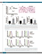
Figure 3. Plt Slc35a1–/– mice exhibit thrombocytopenia. (A) Peripheral blood platelet counts and mean platelet volume in wild-type (WT) (n=10) and Plt Slc35a1–/– (n=10) mice. Each circle or triangle represents one mouse. *** P<0.001. (B) Wright-Giemsa staining of peripheral blood smears from WT (n=3) and Plt Slc35a1–/– (n=3) mice. Platelets are indicated by arrows. Scale bar, 16 mm. (C) Plasma thrombopoietin levels of WT (n=5) and Plt Slc35a1–/– (n=5) mice measured by enzyme- linked immunosorbent assay. Data are means ± standard deviation (SD). **P<0.01. (D) Flow cytometry analysis of the percentage of reticulated platelets stained by anti-CD41 antibody and thiazole orange (TO) in WT (n=12) and Plt Slc35a1–/– (n=12) mice. Data are means ± SD. ***P<0.001. (E) Reticulated platelet counts of WT (n=9) and Plt Slc35a1–/– (n=9) mice after staining with anti-CD41 antibody and thiazole orange. Data are means ± SD. *P<0.05. MPV: mean platelet volume; TPO: thrombopoietin.
A
B
Figure 4. Slc35a1–/– platelets express low levels of GPIba. (A) Wild-type (WT) and Slc35a1–/– platelets isolated from peripheral blood were stained with antibodies to CD42b, CD41 and CD61, and then analyzed by flow cytometry for expression of GPIba, GPIIb-IIIa complex (n=4 for WT and Slc35a1–/– mice, respectively). (B) Megakaryocytes were isolated from WT or Slc35a1–/– bone marrow, and were then stained with antibodies to CD42b, CD41 and CD61. The stained samples were analyzed by flow cytometry for expression of GPIba and GPIIb-IIIa complex (n=3 for WT and Slc35a1–/– mice, respectively).
764
haematologica | 2021; 106(3)
