
Page 116 - 2021_03-Haematologica-web
P. 116
X. Ma et al.
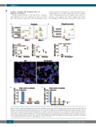
Slc35a1–/– platelets and megakaryocytes are deficient in sialylation
The glycosylation profiles of WT and Slc35a1–/– platelets and megakaryocytes were analyzed by flow cytometry and confocal microscopy based on lectin staining. For the
AB
lectin-based flow cytometry, we used biotinylated lectins, which enabled us to use PE-streptavidin only as a negative control. Neuraminidase-treated samples were used as a positive control (Figure 2A, Online Supplementary Figure S2). Binding of SNA (specific for a2,6-sialic acid) was not
C
DE
Figure 2. Slc35a1–/– platelets and megakaryocytes have reduced sialylation. (A) Top, a representative histogram of flow cytometry analysis of wild-type (WT) and Slc35a1–/– platelets stained with biotinylated RCA 1 (specific for non-reducing terminal β-galactose), or biotinylated MAL II (specific for a2,3-linked sialic acid), and FITC-labeled anti-CD41 antibody. Sialidase-treated platelets were used as a positive control. Platelets treated with phosphate-buffered saline (PBS) were used as a negative control. Unstained, incubation with PE-streptavidin only as a negative control; Bottom, mean fluorescence intensity ratios of RCA 1 versus CD41 or MAL II versus CD41 (n=4). (B) A representative histogram of flow cytometry analysis as shown in (A) on primary bone marrow megakaryocytes (n=4). (C) Representative con- focal microscopic images of WT (n=3) and Slc35a1–/– (n=3) bone marrow megakaryocytes. Arrows, megakaryocytes. Scale bar, 10 mm. (D and E) Ratio of molecular species of sialic acids between WT or Slc35a1–/– platelets. NeuGc, a major isoform of sialic acids in mice. NeuAc, a minor isoform of sialic acids in mice. The numbers 0 – 4 in (E) indicate the number of NeuGc on complex N-glycans. *P<0.05; ***P<0.001.
762
haematologica | 2021; 106(3)
