
Page 145 - 2021_02-Haematologica-web
P. 145
Alternative polarization induces tissue factor
CD206 expression and regions with low TF expression were predominantly negative or low for CD206 (Figure 6C). Besides tumors, atherosclerotic plaques also contain areas of high TF expression.10 We, therefore, analyzed the presence of TF in CD206+ regions or CD80+ regions within human atherosclerotic lesions. We were able to detect TF in both CD206+ regions and in CD80+ regions. However, adjusted TF intensity was higher in regions positive for CD206, indicating an association of increased TF presence close to alternatively activated macrophages (Figure 6D). Furthermore, using flow cytometry, we analyzed macrophages isolated from ath- erosclerotic plaques from ApoE-/- mice fed a high fat diet for 20 weeks. The gating strategy is shown in Online Supplementary Figure S3. Overall, proinflammatory CD80high macrophages were less positive for TF surface expression as compared to CD206high macrophages (Figure 6E). Furthermore, CD206high macrophages had a two-fold higher TF surface intensity staining as deter- mined by a comparison of mean fluorescence intensity of
CD206high macrophages and CD80high macrophages (Figure 6F).
Discussion
Based on extensive experimental work and clinical data it has been well established that circulating monocytes are an important source of TF and, that by expressing this major component of coagulation, they play a key role in linking inflammation and thrombosis in various patholo- gies.10 Much less is known about the expression of TF and its regulation in macrophages. Here, for the first time we provide evidence that in human macrophages the expres- sion of TF is not altered in a proinflammatory environment but is significantly enhanced when these cells are alterna- tively polarized. When human macrophages were polar- ized with IL-4 and IL-13 a significant increase in mRNA specific for TF was observed after 2 h and 6 h. Alternative polarization also caused a significant increase in TF protein
AB
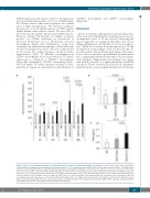
Figure 5. Influence of polarization conditions on extracellular vesicle production of human macrophages. (A) Numbers of extracellular vesicles in the supernatant of unpolarized macrophages and macrophages polarized in the presence of 100 ng/mL lipopolysaccharide (LPS) and 100 ng/mL interferon (IFN)-γ or 20 ng/mL inter- leukin (IL)-4 and 20 ng/mL IL-13 were determined by flow cytometry as indicated in the Methods section. Values are given as total vesicle count per mL: n=9 for 4 h and 12 h; n=12 for 24 h; and n=14 for 48 h. (B) Phosphatidylserine (PS) content of extracellular vesicles in the supernatant of M0 and macrophages polarized in the presence of 100 ng/mL LPS and 100 ng/mL IFN-γ or 20 ng/mL IL-4 and 20 ng/mL IL-13 for 48 h was determined using a specific enzyme-linked immunosorbent assay as indicated in the Methods. Values are given in nM PS (n=6). (C) Total extracellular vesicle-derived RNA was evaluated in the supernatant of M0 and macrophages polarized in the presence of 100 ng/mL LPS and 100 ng/mL IFN-γ or 20 ng/mL IL-4 and 20 ng/mL IL-13 for 48 h as indicated in the Methods section. Values are given in ng/mL RNA (n=5) and represent mean values ± standard deviation. M0: unpolarized macrophages; M(LPS+IFN): classically activated polarized macrophages; M(IL-4+IL13): alternatively activated polarized macrophages.
C
haematologica | 2021; 106(2)
459
