
Page 143 - 2021_02-Haematologica-web
P. 143
Alternative polarization induces tissue factor
coated area. Our results indicate that filipodia of macrophages of classically polarized macrophages M(LPS+IFN) were not affected by the laminin 5 coating whereas in alternatively polarized macrophages, M(IL4+IL13), a significant reduction of filopodia was seen under these conditions (Figure 1D). Interestingly, the inte- grin described for the laminin 5 effect, namely integrin α3 was also downregulated in proinflammatory macrophages (Online Supplementary Figure S1D).
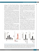
A significant induction of TF mRNA after IL-4 and IL-13 polarization in human macrophages was seen after 2 h and 6 h of stimulation (Figure 2A). To further control for the influence of proinflammatory polarization on TF mRNA, we also performed a time course for TF mRNA levels in M(LPS+IFN). No significant regulation of TF mRNA was observed in proinflammatory macrophages at 2 h (2.9±4.2 fold the level in M0, P=0.5 [n=3]), 6 h (0.9±0.4 fold the level in M0, P=0.8 [n=3]) and 24 h (1.2±0.7 fold the level in M0, P=0.6 [n=3]). The main signaling pathway observed for IL- 4 and IL-13 is an activation cascade dependent on signal transducer and activator of transcription (STAT) 6 signal- ing, as indicated by the increase of phosphorylated STAT6 in macrophages after treatment with IL-4 and IL-13 (Figure 2B). When a STAT6 inhibitor was used, the increase in TF- specific mRNA induced by IL-4 and IL-13 was completely inhibited in these cells (Figure 2C). Blocking poly ADP ribose polymerase (PARP), a possible STAT6 downstream target, also abrogated the IL-4 and IL-13-induced TF expression in these cells (Figure 2D).
Macrophages can be differentiated from monocytes via macrophage colony-stimulating factor (MCSF) or via gran- ulocyte-macrophage colony-stimulating factor (GMCSF). Baseline extracellular vesicle TF levels were higher in GMCSF-derived macrophages than in MCSF-derived macrophages (Figure 3A). However, as can be seen from Figure 3B, when macrophages generated from monocytes with either MCSF or GMCSF were polarized with IL-4 and IL-13, these cells produced significantly more TF than the
respective M0 macrophages and the respective macrophages polarized with LPS and IFN-γ. Polarization of macrophages to M(LPS+IFN-γ) and M(IL-4+IL-13) is in part reversible but has the potential to change subsequent behavior of already polarized macrophages. TF expression was again significantly induced in macrophages by IL-4 and IL-13 in both previous M0 and M(LPS+IFN-γ). Interestingly, previous M(IL-4+IL-13) polarization prevent- ed a significant increase in TF protein after restimulation (Figure 3C).
A
BCD
In contrast to M(LPS+IFN-γ), monocytes treated with LPS and IFN-γ showed significant upregulation of TF (Figure 4A). We found greater methylation around the nuclear factor-kappa B (NF-κB) response element within the TF promoter region in macrophages compared to monocytes obtained from the same donors (Figure 4B). When a chemical demethylating agent23 was used during polarization, TF was induced by LPS and IFN-γ in macrophages, whereas the increase in TF was similar in macrophages polarized with IL-4 and IL-13 and the demethylating agent and in macrophages treated with only IL-4 and IL-13 (Figure 4C).
TF is associated with extracellular vesicles. To under- stand the effect of polarization on extracellular vesicles more globally we determined the changes in extracellular vesicle production in unpolarized and polarized macrophages. As can be seen from Figure 5A, alternative polarization of macrophages using IL-4 and IL-13 resulted in a significant increase of extracellular vesicle production over time when compared to extracellular vesicle produc- tion by unpolarized macrophages. In contrast, proinflam- matory polarization of macrophages with LPS and IFN-γ did not have a significant effect on the production of extra- cellular vesicles. Similar results were obtained using an ELISA, which specifically recognizes phosphatidylserine present on extracellular vesicles (Figure 5B). In addition, we used extracellular vesicle RNA as a surrogate marker for the amount of circulating vesicles. The total amount of
Figure 2. Tissue factor-specific mRNA induction by alternative polarization. (A) Tissue factor (TF) mRNA levels at the indicated time points in macrophages after polarization induced by interleukin (IL)-4 and IL-13 (n=3). (B) Phosphorylated STAT6 as determined in macrophages 30 min after IL-4+IL-13-induced polarization in comparison to that in unpolarized M0 macrophages using flow cytometry and specific antibodies as described in the Methods (n=3). A representative image is shown. (C) TF mRNA levels in macrophages 2 h after IL-4+IL-13-induced polarization in the presence and absence of a specific STAT6-inhibitor (S6Inh.) at 250 mM (n=6). (D) TF mRNA levels in macrophages 2 h after IL-4+IL-13-induced polarization in the presence and absence of the PARP-inhibitor PJ34 at 100 mM (n=6). TF mRNA in panels A, C and D was determined by quantitative polymerase chain reaction and GAPDH was used as a housekeeping gene as indicated in the Methods section. Values are given as fold changes compared to the respective unpolarized control (M0) and represent mean values ± standard deviation.
haematologica | 2021; 106(2)
457
