
Page 65 - 2020_09-Haematologica-web
P. 65
XPO1 is a target to treat β-thalassemia
Here, we validate these previous findings using confocal microscopy and ImageStream analysis for a more quan- titative approach. In normal culture conditions of cord blood-derived CD36+ erythroid progenitors at day 3, a 2- hour treatment with LMB at 20nM induced an increase in HSP70 nuclear accumulation (Online Supplementary Figure S1A and B) resulting in an increase in HSP70 nuclear/cytoplasmic ratio.
In order to determine which specific exportin was involved in HSP70 export, we first analyzed expression of the seven exportins during erythropoiesis. All of them were expressed at the mRNA and protein levels during human erythroid differentiation as shown by transcrip- tomic and proteomic analysis10,11 (Figure 1A and B).
Based on the proteomic database (Figure 1B), we found that exportin-2 (XPO2) is the most expressed exportin at the protein level in human erythroid progenitors. However, since primary function of XPO2 is to mediate re-export of importin-a from the nucleus to the cyto- plasm, it is unlikely to be the candidate exportin involved in HSP70 nuclear export. In contrast, XPO1, in addition to exporting RNAs, mediates export of a broad range of cargo proteins bearing a leucine-rich Nuclear Export Sequence (NES),12,13 that include a large variety of tumor suppressor proteins (e.g. p53, p21, FOXO) and thereby fulfills all the criteria to be a good candidate in exporting HSP70. We performed in silico analysis of published data for GATA-1 chromatin immuno-precipitation (ChIP) in
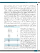
Table 1. GATA-1 chromatin immunoprecipitation (ChIP) peak scores in XPO1 gene and three known erythroid genes.
human erythroid cells,14 and found twelve annotated peaks for GATA-1 in the promoter of the XPO1 gene sug- gesting that it might be an erythroid regulated gene (Table 1). In addition, XPO1 was the second most expressed exportin in erythroblasts, and concomitantly with HSP70 nuclear accumulation, its expression was down-regulated both at the mRNA and protein levels along terminal differentiation (black line in Figure 1A and B). This hypothesis was further supported by in silico analysis showing that human HSP70 protein sequence contains a putative leucine-rich NES at position L394- L400 that could interact with XPO1 (Figure 1C, NES human HSP70 protein WT).
In order to validate this hypothesis, we first showed that XPO1 interacted with HSP70 in vitro by protein-pro- tein interaction experiments using BioLayer interferome- try (BLI) (Figure 1C, red line). Furthermore, we observed a decreased binding affinity between XPO1 and HSP70 with the mutant Serine 400 Alanine (HSP70S400A) in the putative leucine-rich NES (Figure 1C, blue line). This mutant HSP70 shows restricted nuclear localization when transduced in erythroid cells.3 Thus, these results validate the importance of putative NES in the interac- tion of HSP70 with XPO1, and in the cellular localization of HSP70. Next, using proximity ligation assay (PLA/Duolink assay) (Figure 1D) and performing CoIP experiments in normal conditions (Figure 1E), we obtained additional evidence that this interaction is direct and occurs in vivo in CD36+ derived erythroid pro- genitors. Interestingly, the fraction of XPO1 protein immunoprecipitating with HSP70 upon EPO deprivation is increased as shown by CoIP experiments (data not shown).
XPO1 is the exporter of HSP70 in human erythroid progenitors
To document the functional role of XPO1 in human ery- thropoiesis, and its putative target to treat β-TM, we first repressed XPO1 expression in human β-TM erythroblasts. For this purpose, CD36+ cells from a β-TM patient were transduced with lentiviruses expressing a XPO1 specific shRNA (shXPO1) or a sh scramble used as control (shCTL). Two days after transduction, ImageStream quantification showed that the cells transduced with shXPO1 efficiently repressed the expression of XPO1 [mean pixel nuclear XPO1 536.4±3.517 (shCTL) vs. 520.0±2.767 (shXPO1) (P<0.0001] (Figure 1F and Online Supplementary Figure 2A). Similar results were obtained in erythroblasts from cord blood and peripheral blood stem cells (data not shown). In agreement with our hypothesis, XPO1 repression was associated with a significant increase in HSP70 nuclear translocation [similarity score 1.090±0.012 for shCTL vs. 1.378±0.011 for shXPO1 (P<0.0001)] and in HSP70 nuclear accumulation [mean pixel nuclear HSP70 139.1±1.176 for shCTL vs. 159.2±1.173 for shXPO1 (P<0.0001), correspon- ding to a 14.5% increase] (Figure 1F). Similar results were obtained using two different shRNA specifically targeting XPO1. As a control of efficacy, P53 that is a well-known target of XPO1, was also significantly increased in the nucleus (Online Supplementary Figure 2B and C). Taken together, these data suggest that in human erythroid cells, HSP70 interacts with XPO1 through a leucine-rich NES region, which is required for its nuclear export and may thus be a good target to improve ineffective erythropoiesis of β-TM.
Gene name
XPO1
Genomic peak location
Intragenic intron Intragenic intron Intragenic intron Intragenic intron Intragenic intron Immediate down. Intron Promoter
Enhancer
Intergenic
Intergenic
Intergenic
Intergenic
Intragenic exon Immediate down. Intron Enhancer
Enhancer Enhancer Promoter Promoter Enhancer Enhancer Promoter
Peak score
53 695.35 76.79 94.13 231.83 62.86 292.11 110.24 66.87 288.01 73.56 89.17 101.5 3100 72.94 186.49 55.94 898.4 105.72 1259.62 313.4 55.51
EPOR
HBA1
HBA2
Annotated peaks extracted from GATA-1 ChIP public data on human erythroid pro- genitors14 analyzed with Galaxy software.Peak scores and locations for XPO1 and the known erythroid genes EPOR (erythropoietin receptor),HBA1 (Hemoglobin a-1) and HBA2 (Hemoglobin a-2).Peak score in MACS output corresponds to -10*log10pvalue of peak region. P-value corresponds to the probability that the candidate peak (reads enrichment) is random.
haematologica | 2020; 105(9)
2243
