
Page 64 - 2020_09-Haematologica-web
P. 64
F. Guillem et al.
A
B
CD
F
E
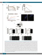
Figure 1 (previous page). In human erythroid progenitors, HSP70 is exported from the nucleus by an XPO1-dependent mechanism. Expression profiles for the seven different exportins (XPO1-XPO7) during terminal erythroid differentiation in human: mRNA expression from proerythroblast stage (ProE) to orthochromatic stage (Ortho). Values are extracted from public data and presented as log of reads per kilobase of transcript per million reads (logRPKM); EB: early basophile; LB: late basophile; Poly: polychromatophilic, (A) and protein expression from progenitor stage (ProG) to orthochromatic stage (Ortho). Values are presented as mean of protein copies per cell. ProG1: BFU-E; ProG2: CFU- E; Baso1: early basophile; Baso2: late basophile; Poly: polychromatophilic (B). Data are representative of three independent experiments. (C) Putative XPO1 specific leucin-rich NES in the protein sequence of human HSP70 (NP_005336) at position L394-L403. The interactions between purified XPO1 and WT HSP70 as well as XPO1 with the nuclear- targeted HSP70 mutant (S400A) were analyzed using BLI. WT HSP70 exhibits a much higher signal (i.e. affinity) for the ligand XPO1 compared to the nuclear HSP70 protein bearing a mutation in the NES residue S400A. Data are representative of two independent experiments. (D) Proximity of HSP70 and XPO1 proteins was analyzed in CD36+ erythroid progenitors derived from cord blood, by Duolink assay, using anti-XPO1 and anti-HSP70 antibodies (or anti-GATA1 for negative control). Red spots indicate <40 nm proximity between cellular-bound antibodies. Nuclei are stained with DAPI (blue). Images have been observed by confocal microscopy (x63 oil objective, scale bar= 5 μm). Data are representative of three independent experiments. (E) HSP70 and XPO1 direct interaction was demonstrated by CoIP experiments. HSP70 and XPO1 immunoblot detection is shown in total lysate (TL), in eluate from HSP70 IP and from IgG Control (IgG CTL) IP. The data are representative of three independent experiments in human erythroïd cells. (F) Erythroid progenitors from β-thalassemia major (β-TM) patient at day 2 of CD36+ culture were transduced with a shRNA specific for XPO1 or a sh scramble (shCTL). Both constructions express GFP. GFP+ cells were sorted and stained with anti-HSP70 or anti-XPO1 antibodies, and DAPI. XPO1 and HSP70 nuclear expression (mean pixel) were analyzed at day 2 following transduction, by ImageStream. In addition, HSP70 nuclear translocation was evaluated by measuring the similarity score between HSP70 and DAPI nuclear stainings. Data are presented as mean ±standard error of mean (SEM). On average, 30,000 events were collected in all experiments. P-values are determined by paired t-test. ***P<0.0001. Three illustrative images (ImageStream) of shCTL and shXPO1 conditions are presented. Cells were probed for HSP70 expression and run on the ImageStream. Bright field (white), DAPI (purple), HSP70 (green), and HSP70/DAPI composite (scale bar=7 μm). Data are representative of six independent experiments, n=2 different β-TM patients with n=2 different shRNA XPO1.
2242
haematologica | 2020; 105(9)
