
Page 43 - 2019_03-Haematologica-web
P. 43
IgG4-related disease for hematologists
CD68+ S100+ histiocytes, often associated with emperipolesis. The most recent classification of the histio- cyte disorders recommends evaluating suspected cases of Rosai-Dorfman-Destombes disease for increased IgG4+
plasma cells,48 but in the absence of other evidence for a common pathophysiological link, Rosai-Dorfman- Destombes disease is not considered part of the spectrum of IgG4-RD or vice versa.49 One third of patients with
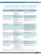
Table 2. Diseases that mimic hematologic manifestations of IgG4-RD (lymphadenopathy, eosinophilia and polyclonal hypergammaglobulinemia).
Mimicker of IgG4-related disease
Multicentric Castleman disease (MCD)
Cutaneous and systemic plasmacytosis (CSP)
Rosai-Dorfman-Destombes disease (RDD, a.k.a “Sinus Histiocytosis with Massive Lymphadenopathy; “R” group histiocytosis)
Erdheim Chester disease (ECD; “L” group histiocytosis)
Malignant lymphoma
Sarcoidosis
Hypereosinophilic syndrome (HES)/chronic eosinophilic leukemia (CEL)
[particularly lymphocyte-variant HES]
Plasma cell myeloma/monoclonal gammopathy of undetermined significance
Vasculitis, particularly eosinophilic granulomatosis with polyangiitis and granulomatosis with polyangiitis
Areas of overlap
• Lymphadenopathy (particularly the MCD-like variant of IgG4-LAD)
• High serum IgG4
• IgG4+ plasma cells in tissue*
• Polyclonal hypergammaglobulinemia
• Lymphadenopathy
• Polyclonal hypergammaglobulinemia
• Polyclonal plasmacytosis, including IgG4+
plasma cells in bone marrow and skin*
• Lymphadenopathy
• Extranodal RDD can involve the nasal cavity,
and retro-orbital tissue
• Meningeal involvement of RDD may mimic
IgG4-pachymeningitis
• Salivary gland involvement
• CNS involvement including pachymeningitis • Increased IgG4+ plasma cells in tissue*
• Retroperitoneal fibrosis
• Central nervous system/hypophysitis • Pulmonary involvement
• Lymphadenopathy
• Extranodal mass lesions
• Blood and tissue eosinophilia
• Lymphadenopathy
• Pulmonary nodules
• Pachymeningitis and/or hypophysitis • Polyclonal hypergammaglobulinemia • Multi-organ involvement
• T-cell clonality by PCR
• Atopy/asthma/elevated IgE
• Lymphadenopathy
• Eosinophilia
• Elevated serum IgG4
• Aberrant T cell phenotype in peripheral
blood (CD4+/3-, CD4+/7–, CD3+/4–/8–)
• Plasma cell infiltrate
• Hypergammaglobulinemia • Renal failure
• Proteinuria
• Eosinophilia in blood and tissue
• Polyclonal hypergammaglobulinemia with
elevated serum IgG4
• Peri-aortitis and rarely, Kawasaki-like coronary
arteritis are seen in IgG4-RD
• Multi-organ involvement, including respiratory,
gastrointestinal and renal structures
Distinguishing features of the mimicker not typically seen in IgG4-related disease
• MCD is a “hyper-IL-6” syndrome associated with
B symptoms and highly elevated CRP and IL-6 not seen in IgG4-RD
• IgG4-RD rarely involves skin whereas cutaneous lesions (round/oval, red/brown poorly circumscribed macules, papules and plaques) are an obligatory feature of CSP; serum IgG4 in CSP is normal or mildly elevated
• Massive cervical lymphadenopathy is atypical for IgG4-RD • RDD may present with B symptoms
• Large histiocytic cells with hypochromatic nuclei and
pale cytoplasm; emperipolesis; positive for S100, CD68, CD14 and CD163
• >95% of ECD patients have bone involvement, which is rare in IgG4-RD (apart from rare IgG4+ angiocentric eosinophilic fibrosis of the head and neck)
• Yellow peri-orbital xanthelasmas common in ECD (skin involvement in IgG4-RD is rare, and when present tends to be erythematous or flesh-colored papules)
• Foamy multi-nucleated histiocytes, few Touton cells, fibrosis; CD68+, CD163+, CD1a–
• 50% of ECD patients are BRAF V600E-positive
• B symptoms, bone involvement, brain parenchymal involvement and hypercalcemia are rare in IgG4-RD
• Non-caseating granulomas
• Hypercalcemia and elevated ACE levels
• Increased blasts or myeloid clone in CEL
• PDGFR-alpha/PDGFR-beta/FGFR1/PCM1-JAK2 positivity
are not seen in IgG4-RD
• More marked and persistent eosinophilia in HES
• Plasma cell clonality
• Monoclonal paraprotein ± suppression of polyclonal
immunoglobulins
• Lytic boney disease/hypercalcemia
• Light chains in urine rather than albuminuria
• Highly elevated CRP in vasculitis
• Extravascular granulomas, small-medium vessel vasculitis • Mononeuritis multiplex not a feature of IgG4-RD
*Although increased IgG4+ plasma cells have been described in these diseases, the absolute counts and IgG4/IgG ratio are typically well below the thresholds for the diagnosis of IgG4- RDandtheotherkeyfeaturesofIgG4-RD(storiformfibrosisandobliterativephlebitis)arenotseen.3 IgG4-LAD:IgG4-lymphadenopathy;IL-6:interleukin-6;CRP:C-reactiveprotein;IgG4- RD: IgG4-related disease; CNS: central nervous system; ACE: angiotensin-converting enzyme; PCR: polymerase chain reaction.
haematologica | 2019; 104(3)
449
