
Page 45 - 2019_03-Haematologica-web
P. 45
IgG4-related disease for hematologists
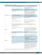
Table 3. Diagnostic and staging tests in IgG4-RD. Test(s)
Complete blood count/differential/blood film
C-reactive protein (CRP); interleukin-6 and other markers of systemic inflammation
Serum IgG subclasses
Immunoglobulins (IgG, IgA, IgM, IgE); serum and urine protein electrophoresis (SPEP, UPEP)
Autoantibodies (e.g. antinuclear antibody, rheumatoid factor)
Complements (C3/C4)
Urinalysis, random albumin/creatinine ratio
Other markers of end organ damage: lipase, glucose and hemoglobin A1c, liver enzymes, thyroid-stimulating hormone, creatinine, urinalysis, urine albumin/creatinine ratio
Computed tomography (CT) of the neck, chest, abdomen, pelvis
If lacrimal enlargement ⇒CT to head (rule out orbital involvement)
Archived specimens
New biopsy
Lymph node and bone marrow
Typical findings
Eosinophilia (~40% of patients, typically mild); rouleaux formation due to polyclonal hypergammaglobulinemia
Normal or moderately elevated (CRP typically <20 mg/L) in the absence of peri-aortitis or active infection
Mildly elevated IgG4 <1.5-5 g/L is nonspecific, and 30% of IgG4-RD patients have normal serum IgG4 levels. Serum IgG4 >5 g/L is helpful both for diagnosis and as a disease marker. Other IgG subclasses may be moderately elevated (IgG4/IgG ratio is typically >0.2)*
IgA and IgM may be normal or mildly increased; IgE may be markedly increased. Immunoglobulin suppression is atypical. SPEP and UPEP are important to rule out monoclonal proteins
May be weakly positive
Often low, especially with tubulointerstitial nephritis
Albuminuria is common; nephrotic range proteinuria can be seen with membranoproliferative glomerulonephritis
Subclinical pancreatitis with elevated lipase, glucose intolerance, hepatitis and albuminuria are common
Diffuse “sausage-like” or segmental enlargement of pancreas, often with a “halo”; wedge-shaped hypodensities in kidneys; ductal organs, such as bile duct and bronchus, show diffuse “pipe-stem” wall thickening; thickened aortic wall; hepatic mass lesions; retroperitoneal fibrosis or peri-aortic cuffing
Patients with orbital disease typically have lacrimal
gland swelling
As long as tissue blocks are still available, the pathologist should be able to examine the histology and then order immunostaining for IgG4 and IgG if typical features are present
Excisional is preferable to core biopsy when possible to look for the central pathological features:
• Storiform fibrosis
• Obliterative phlebitis
• Polyclonal lymphoplasmacytic infiltrate with increased
IgG4+ plasma cells and IgG4+/IgG+ plasma cell ratio > 40%**
Consider minor salivary gland (lip) biopsy if affected organs
are high risk for biopsy
These tissues are unusual in that fibrosis and obliterative phlebitis are typically not seen, and thus biopsy of other tissues may be required for a definitive diagnosis. Involved lymph nodes typically have >100 IgG4+ plasma cells/hpf
with an IgG4/IgG ratio >40%. Bone marrow involvement with eosinophilia and increased IgG4+ plasma cells is rare and may be absent even in patients with marked serum hypergammaglobulinemia
Imaging
Pathology
*IgG2 may be spuriously elevated when nephelometric measurement is used.65 **The number of IgG4+ cells/hpf required varies, depending on the tissue, from >10/hpf in meningeal tissue to >200/hpf in skin. In all tissues, the ratio of IgG4+ to IgG+ plasma cells should be ≥40%.3 Hpf: high-power field.
haematologica | 2019; 104(3)
451
Blood and urine tests
