
Page 46 - 2019_03-Haematologica-web
P. 46
L.Y.C. Chen et al.
cytic infiltrate, the number of IgG4+ plasma cells per high- power field (hpf) considered diagnostic varies according to tissue site, from >10/hpf in meninges to >100/hpf in skin. Regardless of the site, the ratio of IgG4+/IgG+ plasma cells is >40% in IgG4-RD. Fibrosis is a histological requirement for the diagnosis of IgG4-RD and should be arranged at least focally in a storiform pattern. Storiform fibrosis is a swirling, “cartwheel” pattern of fibrosis which may have a patchy distribution and can, therefore, be missed with small biopsies. In the obliterative phlebitis of IgG4-RD, venous channels are obliterated by an inflammatory lym- phoplasmacytic infiltrate. Expert pathologists recommend looking for arteries/arterioles where the accompanying venous vessel is not readily apparent and may in fact have been replaced by an inflammatory infiltrate; elastin stains may be helpful in identifying completely obliterated ves- sels.
Other histopathological features include phlebitis with- out obliteration of the lumen and increased number of eosinophils. As in the illustrative case, archival specimens may be used to confirm a diagnosis, and many patients will have previous biopsies available due to their chronic disease course. As long as a tissue block is still available, IgG4 and IgG stains can be done. When a new biopsy is required, excisional specimens are preferred over core nee- dle biopsies to allow for proper assessment. There are cur- rently no established criteria for the cytological diagnosis of IgG4-RD from a fine needle aspirate.
Increased numbers of IgG4+ plasma cells are seen in all tissues affected by IgG4-RD, but this is not, in itself, a spe- cific finding. Many chronic inflammatory conditions such as vasculitis, inflammatory bowel disease and lymphoma may exhibit increased numbers of IgG4+ plasma cells but do not share the other histological features of storiform fibrosis, obliterative phlebitis and absence of granuloma- tous inflammation.19,21 Unfortunately, for the purposes of hematologists, bone marrow involvement is uncommon in IgG4-RD and obliterative phlebitis and storiform fibro-
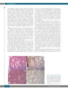
sis (not to be confused with myelofibrosis) are not typical- ly seen in bone marrow and lymph nodes.70 Furthermore, even when involved, lymph nodes and bone marrow may not show robust elevation in IgG4-expressing plasma cells as compared to the total IgG population, or the findings may be only focal. Patterns of IgG4-RD have not been well established in the bone marrow, but the presence of mature plasma cells would be supportive of marrow involvement, once plasma cell neoplasms are excluded. Some examples of marrow involvement in IgG4-RD are shown in Figure 4.
In general, only organs with clinical or radiological evi- dence of involvement are likely to show diagnostic fea- tures on biopsy, and thus biopsy should be directed at affected organ(s). Patients with proteinuria or renal lesions on imaging may require kidney biopsy, and renal involve- ment may demonstrate two distinct histological patterns: hypocomplementic tubulointerstitial nephritis in 80% of cases, and membranoproliferative glomerulonephritis in 20% of cases.70 In patients in whom affected organs are not amenable to biopsy, minor salivary gland (lip) biopsy should be considered. Even without clinical evidence of major salivary gland swelling or sicca symptoms, minor salivary gland biopsy can be a minimally invasive way to reach a histological diagnosis in some patients. One study of 66 patients with suspected IgG4-RD reported a sensitiv- ity of 55% and specificity of 100% for labial salivary gland biopsy.71 From a pragmatic perspective, patients who have classic clinical, laboratory and radiological manifestations of IgG4-RD but are too frail to undergo an attempted biopsy attempt, or in whom small biopsies yield insuffi- cient diagnostic material,72 can be given a working diagno- sis of “suspected IgG4-RD” and treated as such provided reasonable efforts have been made to exclude mimickers of IgG4-RD.
Splenic involvement in IgG4-RD remains an enigma. Overt splenomegaly and splenic lesions are rare in con- firmed cases of IgG4-RD. A rare entity known as scleros-
AB
CD
Figure 4. Bone marrow specimens involved by IgG4-related disease. Both cases show mature plasma cells distrib- uted throughout the marrow. Ancillary studies established that these plasma cells were polyclonal, excluding a plasma cell neoplasm. (A,C) Hematoxylin and eosin stains. (B,D) IgG4 immunohisto- chemistry.
452
haematologica | 2019; 104(3)
