
Page 42 - 2019_03-Haematologica-web
P. 42
L.Y.C. Chen et al.
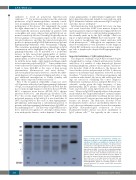
sufficient to result in polyclonal hyperviscosity syndrome.27,28,34 It is not known what causes the exuberant production of IgG4 immunoglobulins, currently consid- ered an epiphenomenon rather than a contributor to the pathogenesis of the disease.8 Not surprisingly, the serum free light chains tend to be abnormally elevated.35 IgE is often markedly increased, particularly in patients with eosinophilia and atopy, whereas IgA and IgM levels are normal or modestly elevated. Serum IgG4 typically runs in the fast gamma or beta-gamma region on the serum pro- tein electrophoresis, and thus the typical electrophoretic profile of a patient with IgG4-RD demonstrates polyclonal hypergammaglobulinemia with beta-gamma bridging. This sometimes prominent pattern is dependent on IgG4 concentration and is highlighted in Figure 3. The hyper- gammaglobulinemia can be mistaken for a polyclonal increase in IgA, monoclonal gammopathy of undeter- mined significance or “biclonal” IgG kappa and lambda gammopathy, as laboratory physicians may not be famil- iar with the dense bands of IgG-lambda and kappa which in fact represent polyclonal IgG4.36,37 Some patients have even been treated for myeloma before subsequently being found to have IgG4-RD as the cause of their protein abnormalities, plasmacytosis and renal disease.9,38 Laboratory physicians must, therefore, consider the differ- ential diagnosis of beta-gamma bridging and order or sug- gest additional investigations to clarify clonality and heavy chain composition where necessary.39,40
Prior to the recognition of IgG4-RD, a large case series of 130 patients with polyclonal hypergammaglobulinemia >30 g/L on serum protein electrophoresis showed that the most common single diagnoses were liver disease (79/130, 66%), connective tissue disease (28/130, 22%), chronic infection (8/130, 6%) and hematologic disorders (7/130, 5%).41 In a recent, single-center study of 70 patients with polyclonal increases in IgG ≥20 g/L it was found that 14 (20%) had IgG4-RD as the cause of their hypergamma- globulinemia, indicating that a substantial proportion of patients with hypergammaglobulinemia have IgG4-RD as the underlying cause.42 The discovery of IgG4-RD has also led to increased recognition of other IgG subclass eleva- tions with specific diseases, such as hepatitis C and mono-
clonal gammopathy of undetermined significance with IgG1, hypothyroidism and irritable bowel syndrome with IgG2, rheumatoid arthritis with increased IgG3 and IgG1, and celiac disease with IgG4.43
IgG4 myeloma has been described but is rare; one large case series found that 6/158 bone marrow biopsies in myeloma patients expressed IgG4, in keeping with the rel- atively small fraction of overall circulating gamma globu- lins normally made up by the IgG4 subtype.10 One case report of IgG4 subtype POEMS has been reported.44 The bone marrow morphology may mimic myeloma with florid plasmacytosis,9,45 although in our experience, bone marrow examination is very insensitive for the diagnosis of IgG4-RD, with many cases showing no increase in plas- ma cells or lymphocytes despite florid hypergammaglob- ulinemia.
Important mimickers of IgG4-related disease
The diagnostic challenge of IgG4-RD for hematologists is heightened by overlap of clinical and laboratory features with those of a number of other hematologic diseases including lymphoma, plasma cell neoplasms, and histio- cyte disorders (Table 2). In addition to the diseases dis- cussed in this section and presented in Table 2, there are numerous non-hematologic mimickers reviewed in detail elsewhere.46 Careful review of histological specimens and correlation with clinical, laboratory and radiological find- ings are crucial for solidifying the correct diagnosis. Multicentric Castleman disease and IgG4-RD show con- siderable overlap given the high frequency of lym- phadenopathy, IgG4+ plasma cell-enriched tissue infil- trates and elevated serum IgG4 levels seen in both dis- eases.18 However, IgG4-RD typically affects older patients, rarely exhibits the “hyper-interleukin-6” systemic inflam- matory features of multicentric Castleman disease such as fever and elevated C-reactive protein, and the histological features are distinct. The histiocytic disorders Rosai- Dorfman-Destombes disease and Erdheim-Chester dis- ease both cause inflammatory mass lesions that can mimic IgG4-RD. Histopathological evaluation of Rosai- Dorfman-Destombes disease can show enrichment of IgG4+ plasma cells,22,47 but typically in the context of
Figure 3. Serum protein electrophoresis showing elec- trophoretic patterns for four patients with mild to gross elevations in IgG4 concentration in between two for patients with low IgG4 concentration. The physicochemi- cal properties of the IgG4 heavy chain result in a relative anodal position (shift toward albumin) of the gamma glob- ulins when the IgG4 becomes the predominant gamma globulin. Apart from IgG4, IgA immunoglobulins are fre- quently observed in the boundary between the beta and gamma regions. Monoclonal bands may also migrate in this region as shown in the gel (the monoclonal gammopa- thy in this case is an IgG1 monoclonal band that has physicochemical properties that are atypical for IgG1 immunoglobulins, which are normally found in a more cathodal position). NC: normal control, MG: monoclonal gammopathy.
448
haematologica | 2019; 104(3)
