
Page 41 - 2019_03-Haematologica-web
P. 41
IgG4-related disease for hematologists
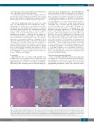
scattered larger or transformed follicles containing plasma cells. An example is given in Figure 2D,E.
(v) Inflammatory pseudotumor-like: lymph node partial- ly effaced by a fibro-inflammatory infiltrate and storiform fibrosis; this subtype is considered most specific for IgG4- RD in lymph nodes. An example is given in Figure 2F.
IgG4-related lymphadenopathy has aptly been called both “an underdiagnosed and overdiagnosed entity”:19 underdiagnosed because if it is not included in the differ- ential diagnosis, IgG4 and IgG stains may not be done and the disease may be missed, and overdiagnosed because increased IgG4+ plasma cells may be seen in conditions ranging from Rosai-Dorfman-Destombes disease to inflammatory vasculitis.21,22 Although not the optimal tis- sue for making the histological diagnosis of IgG4-RD, in a patient with typical clinical features such as autoimmune pancreatitis or retroperitoneal fibrosis, a lymph node biop- sy may be sufficient for diagnosis if biopsy of other affect- ed organs is not feasible. Given the low specificity of increased IgG4+ plasma cells in lymph node and the vari- able histological patterns, the greatest utility of lymph node biopsy is perhaps excluding other diagnoses, such as lymphoma and HHV8-associated Castleman disease. The role of lymph node biopsy is further discussed in the sec- tion on “Diagnosis and staging”.
Eosinophilia
Approximately 40% of patients with IgG4-RD have peripheral blood eosinophilia, often accompanied by asth- ma and atopy.23 Thus, IgG4-RD is an important and under- appreciated cause of reactive or secondary eosinophilia.12 HES and IgG4-RD commonly involve the skin, lungs, gas-
trointestinal tract, and lymph nodes.12 Idiopathic HES and hypereosinophilia of unknown significance are diagnoses of exclusion, and account for a substantial proportion (30- 50%) of diagnoses of patients evaluated for eosinophilia.24- 26 Evaluating these patients for IgG4-RD is an important and underappreciated aspect of their care. In fact, we pre- viously published a case report with a diagnostic label of idiopathic HES, reviewed by several world experts in eosinophilia who concurred with the diagnosis, which was subsequently found to be IgG4-RD.27,28 Findings of a myeloid clonal disorder such as increased blast cells, abnormal karyotype, mutations in PDGFR-alpha/beta, FGFR-1 and PCM1-JAK2 are not seen in IgG4-RD. However, differentiating lymphocytic-variant HES from IgG4-RD can be more challenging. The aberrant T-cell phenotypes found in lymphocytic-variant HES, including increased CD4+CD3–, CD3+/CD4–/CD8– and CD4+/CD7– T cells, with or without T-cell clonality as determined by polymerase chain reaction analysis, have all been reported in IgG4-RD.12,29 Increased IgG4 deposits have been found in tissue samples from adult and pediatric patients with eosinophilic esophagitis.30-33 In contrast to HES and chron- ic eosinophilic leukemia, the eosinophilia secondary to IgG4-RD is typically mild to moderate, rarely exceeding 5x10⁹/L and typically quite evanescent, being ablated by steroids or rituximab therapy.
Polyclonal hypergammaglobulinemia
As for eosinophilia, IgG4-RD represents an important new diagnostic consideration in patients with hypergam- maglobulinemia. An elevated serum IgG4 level, often accompanied by an increase in IgG1, causes polyclonal hypergammaglobulinemia. Rarely, this elevation can be
ABC
DEF
Figure 2. Lymph nodes in IgG4-related disease. (A,B) An example of the interfollicular pattern of IgG4-related lymphadenopathy, with mature plasma cells, many expressing IgG4, distributed between benign follicles. (A) Hematoxylin and eosin stain. (B) IgG4 immunohistochemistry. (C) A needle core lymph node biopsy from a different case with the interfollicular pattern (hematoxylin and eosin stain). (D,E) A case of IgG4-lymphadenoapthy with a progressive transformation of the follicular center pattern, with the plasma cells within the follicle proper. (D) Hematoxylin and eosin stain. (E) IgG4 immunohistochemistry. (F) An example of a mass-like lesion (inflammatory pseudotumor) with dense fibrosis and associated follicular hyperplasia in a case of IgG4-lymphadenoapthy (hematoxylin and eosin).
haematologica | 2019; 104(3)
447
