
Page 209 - 2019_03-Haematologica-web
P. 209
IL6ST variants lead to aberrant T-cell phenotype
the autosomal dominant form of HIES, but capillary sequencing performed prior to whole exome sequencing did not reveal a mutation in STAT3.18 Family history was remarkable for the early death of 3 siblings (Figure 1A). The first female child (III-1 on Figure 1A) had scapho- cephaly, foot deformities, recurrent diarrhea, respiratory infections, keratitis, and retarded growth and develop- ment. She died at the age of 3.5 years due to intestinal per- foration. The second gestation resulted in twin brothers: III-2 was born prematurely at 24 weeks with a foot defor- mity and died 2 h after birth, whereas III-3 died in utero at 20 weeks. The third gestation was PP498L’s older brother (III- 4) who has congenital blindness, and the fourth was PP498L.
Patient PN404Y bearing a GP130N404Y mutation has been described previously.5 The marked similarity of clinical
phenotypes between PP498L and PN404Y is illustrated in Online Supplementary Table S3.
Identification of a novel IL6ST mutation
We performed whole exome sequencing in PP498L to iden-
tify the underlying molecular disease etiology, and identi- fied a homozygous missense mutation in IL6ST (c.1493C>T, p.P498L) (Figure 1D) deemed disease-causing based on functional predictions (Online Supplementary Table S4) and phenotypic similarity with the recently reported IL6ST-mutant (p.N404Y) patient, PN404Y.5 These amino acid positions are within the fifth and fourth domains of GP130, respectively, forming crucial interac- tions with other residues to maintain the acute bend in the
A
B
CD
E
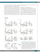
F
Figure 3. Functional assessment of GP130P498L variant in fibroblasts. (A-E) Dose-escalation curves showing relative mean fluorescence intensity (rMFI) of p-STAT3 after stimulation of PP498L and healthy donor (HD) fibrob- lasts as well as PP498L fibroblasts transduced with wild-type (WT) GP130 with (A) IL-6, (B) IL-11, (C) IL-27, (D) LIF, (E) OSM, following overnight star- vation in serum-free media. Bar graphs (right) showing rMFI of fibroblasts upon stimulation with the highest concentration of the corresponding cytokine: 3 (HD), 6 (p.P498L) and 4 (p.P498L + WT GP130) replicates of 3 independent experiments; Mann-Whitney-test; *P<0.05, **P<0.01. (F) Percentage of p-STAT4 assessed in PP498L and HD-derived fibroblasts after stimulation with LIF (100 ng/mL) following a 3-hour starvation in serum- free media. Three independent experiments were performed; the shapes of the symbols represent the individual experiments.
haematologica | 2019; 104(3)
615
