
Page 210 - 2019_03-Haematologica-web
P. 210
T. Shahin et al.
protein structure that is conserved across species (Figure 1E and F).19 We found GP130 protein expressed in PP498L fibroblasts, albeit at lower levels (Figure 1G), and in a CRISPR-engineered HEK293-IL6ST knockout (KO) cell line with transient overexpression of the GP130N404Y mutant.5
Functional assessment of GP130P498L mutation in primary and patient-derived cells
Phosphorylation of STAT3 (p-STAT3) is a direct down- stream effect of GP130 activation. To assess the impact of the p.P498L substitution, we studied STAT3 phosphoryla- tion in primary T cells from PP498L and observed markedly decreased p-STAT3 levels upon stimulation with IL-6 and IL-27 compared to stimulation with IL-10 or IL-21 and to healthy donors (Figure 2A). Our previous study5 on PN404Y showed that IL-6 signaling had been abolished but a smaller reduction in p-STAT3 after stimulating primary T cells with IL-27 (CD3+, CD4+, and CD8+ T cells) (Figure 2B), indicating a mutation-dependent effect on the severity of downstream signaling through selected cytokines. Furthermore, IL-6 sig- naling was shown to be defective in both EBV-LCLs and T lymphoblasts derived from PBMCs of PP498L, and IL-27 (that
stimulates T lymphoblasts) was aberrant in PP498L (Figure 2C and D). In addition, we tested the effect of the new p.P498L substitution on the activation of other STAT family tran- scription factors in T lymphoblasts. Stimulation with IL-6 mainly activated STAT3 in healthy donors with no com- pensatory increase in activation of STAT1 in the patient cells (Online Supplementary Figure S1A), whereas phosphory- lation of STAT1, STAT3 and STAT4 was abolished in patient T lymphoblasts upon stimulation with IL-27 com- pared to healthy donor-derived cells (Online Supplementary Figure S1B). Activation of STATs by GP130-independent cytokines including IL-4, IL-21 and IFNb was unaffected in patient cells, except for STAT4 which showed increased phosphorylation upon IL-12 stimulation of PP498L T lym- phoblasts (Online Supplementary Figure S1C-F).
To further evaluate the spectrum of mutation-dependent signaling defects, we analyzed p-STAT3 responses in fibroblasts from PP498L and a healthy donor, after overnight starvation. IL-6 or IL-11 stimulation demonstrated signifi- cantly reduced p-STAT3 levels (Figure 3A and B), with the aberrant IL-11 signaling likely to underlie the majority of bone manifestations in PP498L.14 OSM stimulation resulted in a partial and statistically significant reduction in p-STAT3
AB
CD
E
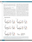
Figure 4. Functional assessment of GP130P498L variant in GP130-KO HEK293 cell line. (A-E) Relative mean fluorescence intensity (rMFI) of p-STAT3 in GP130 CRISPR- knockout HEK293 cells that were transfected with a plasmid coding for the GP130P498L (GP130 KO + p.P498L), wild-type GP130 (GP130 KO + WT GP130) or trans- fected with the empty plasmid (GP130 KO + eV), after stimulation with (A) IL-6, (B) IL-11, (C) IL-27, (D) LIF, (E) OSM. From left to right: dose-escalation curves, stacked histograms displaying shifts in p-STAT3 signals, and bar graphs showing rMFI of fibroblasts upon stimulation with the highest concentration of the corresponding cytokine. (6 replicates of 3 independent experiments are shown; Wilcoxon matched-pairs signed rank test; *P<0.05.)
616
haematologica | 2019; 104(3)
