
Page 207 - 2019_03-Haematologica-web
P. 207
IL6ST variants lead to aberrant T-cell phenotype
Results
Clinical disease manifestation and immunological characterization of Patient PP498L
Patient PP498L was born to consanguineous Turkish par- ents (first-degree cousins) (Figure 1A). He experienced diarrhea at one month of age, recurrent otitis media, bilat- eral keratitis, and recurrent respiratory infections includ- ing pneumonia (Serratia marcescens and Pseudomonas aerug- inosa were isolated) complicated by empyema and pneu- mothorax. In addition, he was followed up for severe eczema and food allergy (milk, egg and wheat; class III).
A
B
At 12 years of age, he developed an aphthous tongue ulcer suggestive of an undefined fungal lesion. No neu- tropenia was recorded, yet he benefited from granulocyte colony stimulating factor. He is currently under monthly intravenous immunoglobulin (IVIG) and cyclosporine A therapy.
Early after birth, the patient exhibited flexion contrac- tures of the hand joints and presented with scaphocephaly (suggesting craniosynostosis) (Figure 1B) and Arnold- Chiari type 1 malformation. He has thoracolumbar scolio- sis (30°) (Figure 1B), clubbing, crowded teeth and mild macroglossia. Cartilage destruction and erosion were seen
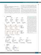
Figure 2. Functional assessment of GP130P498L variant in primary cells. (A) Assessment of GP130 function by flow cytometry measure- ment of percentage of p-STAT3 positive cells after stimulation of primary T cells with IL-6 (100 ng/mL), IL-27 (100 ng/mL), IL-10 (50 ng/mL) or IL-21 (100 ng/mL) in PP498L (orange), mother of PP498L (green), and 4 healthy donors (HD: blue; age-matched HDs are represented by circles and adult HDs by squares). (B) Overlayed histograms showing shifts in p-STAT3 signal upon IL-27 stimulation (solid line) and baseline (dotted lines) in CD3+, CD4+ and CD8+ T cells of both PP498L (orange) and PN404Y (red) compared to a HD (black). Values represent percentage of p-STAT3 positive cells. (C and D) Percentage p-STAT3 assessed in (C) T lym- phoblasts from PP498L and 2 HDs and in (D) Epstein-Barr virus-transformed lymphoblastoid cell lines (EBV-LCLs) from PP498L, mother of PP498L and 2 HDs after stimulation with IL-6 (100 ng/mL), IL-10 (50 ng/mL), IL-21 (100 ng/mL), or IL-27 (100 ng/mL). Data shown are from 2-3 independent experiments (shown by different data point shapes) with 2-3 replicates each. Statistical analysis on IL-6 stimulation of T lym- phoblasts (3 independent experiments) was performed using an unpaired two-tailed Student t-test on the means of the technical replicates. **P<0.01, n=3.
C
D
haematologica | 2019; 104(3)
613
