
Page 144 - 2019_03-Haematologica-web
P. 144
F. Koczian et al.
cells with PS89 in combination with etoposide, vincristine or 6-mercaptopurine, respectively, activation of caspase-3 and PARP cleavage indicated a clear induction of apoptotic cell death (Figure 1C, Online Supplementary Figure S2). In addition, apoptosis was prevented by the pan-caspase inhibitor QVD-OPh (Online Supplementary Figure S3). Since pharmacokinetic studies demonstrated that PS89 has a very short half-life in the blood (data not shown), in vivo
experiments at not feasible at the moment. However, the broad applicability of PS89 as a chemosensitizing agent was confirmed in ALL and AML PDX cells of diverse backgrounds (Online Supplementary Table S2). PDX sam- ples treated with PS89 and vincristine (Figure 1D) or PS89 and daunorubicin (Figure 1E) showed clearly increased apoptosis rates compared to those treated with the single cytostatics (distinct P-values are presented in Online
AB
CD
EF
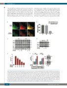
Figure 3. Signal transduction via the BAP31-caspase-8 axis. (A) Immunofluorescence staining of BAP31 (green) and localization of the PS89 photo probe linked to a rhodamine reporter (red) in HeLa cells. Representative original images (upper row) and areas of co-localization analyzed with Leica LAS X software (bottom row) are shown. (B) Diffusion values of PS89 and PS89+BAP31 showing an interaction between the probe and protein. Control samples of 50 nM freely diffusing PS89 in buffer solution and 50 nM PS89 probe plus 50 nM BAP31 protein in buffer solution were analyzed by single-point fluorescence correlation spectroscopy meas- urements. The diffusion coefficient of the probe alone was measured after one-species fitting of the autocorrelation curves (n>15). Diffusion coefficients (D1, D2) of S1 and S2 were measured after two-species fitting of the autocorrelation curves (n>10). The diffusion time (D2) of the mixtures was confined to the diffusion value obtained in the control experiment with PS89 probe alone. Bars represent the mean + standard error of mean. (C) Cleavage of caspase-8 (CASP8) and BAP31 was determined by immunoblotting in Jurkat cells treated with PS89 and etoposide (ETO) for 24 or 48 h. (D) Co-immunoprecipitation of BAP31 and CASP8 from Jurkat cell lysates after 24 h stimulation with PS89 and ETO. Blots were probed for BAP31 and pro- and intermediate p43/41 CASP8. (E) Apoptosis of Jurkat cells treated with the PS89 and ETO combination in the presence of the specific CASP8 inhibitor Z-IETD-FMK after 48 h. (F) Apoptosis of BAP31-silenced HeLa cells treated for 48 h with PS89 and ETO (6 h after transfection). The percentage of apoptotic cells was determined by FACS analysis and normalized to controls. The effect of the PS89 + ETO combination versus treatment with ETO alone was analyzed in siCtrl and siBAP31 cells (one-way ANOVA, Tukey test, P<0.05).
550
haematologica | 2019; 104(3)
