
Page 145 - 2019_03-Haematologica-web
P. 145
Supplementary Table S3A,B). The Bliss independence model confirmed the synergistic effect of PS89 in combination with vincristine or daunorubicin in ALL and AML PDX cells. Nevertheless, in accordance with previous results, PS89 was non-toxic. Notably, compared to ALL and AML patients’ samples, both healthy peripheral blood mononu- clear cells and CD34-positive hematopoietic stem cells showed weak responses to combination treatments (Figure 1D-F, Online Supplementary Table S3).
A
PS89 - a novel option for combination therapy in acute leukemia
Proteomics identified a PS89 target network affecting endoplasmic reticulum homeostasis
To elucidate the mechanisms underlying the impressive pro-apoptotic effect of PS89 combination treatments, the role of the prominent PS89 target, PDI, was studied by genetic knockdown and overexpression experiments. Since PDI silencing did not mimic and PDI overexpression did not rescue the sensitizing effect of PS89 on apoptosis induction or inhibition on proliferation (Figure 2A and
B
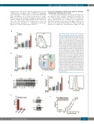
Figure 4. Pro-apoptotic crosstalk at the endoplasmic reticulum-mitochondria interface. (A) Intracellular calci- um levels of Jurkat cells treated with PS89 and etopo- side (ETO) for 24 and 48 h. Fluorescence of Cal-520 stained cells was determined by FACS analysis and mean values normalized towards a dimethyl sulfoxide (DMSO) control. The dotted gray line represents unstained controls. (B) Mitochondrial depolarization of Jurkat cells treated with PS89 and ETO for 24 and 48 h. The percentage of JC-1-stained cells with dissipated ver- sus intact membrane potential (ΔΨm) was determined by FACS analysis (populations as shown by FACS dot plots). (C) Cytochrome c release from mitochondria into the cytosol. Fractionation of Jurkat cell lysates after 48 h treatment with PS89 and ETO was confirmed by volt- age-dependent anion channel (VDAC) immunoblotting. Stain-free gels served as the loading control. (D) Intracellular levels of reactive oxygen species (ROS) in Jurkat cells treated with PS89 and ETO for 24 h and 48 h. Fluorescence of carboxy-H2DCFDA-stained cells was determined by FACS analysis and mean values normal- ized to a dimethyl sulfoxide (DMSO) control. The dotted gray line represents unstained controls. (E) Apoptosis of Jurkat vector control (Jurkat/neo), Bcl-2 overexpressing (Jurkat/Bcl-2) and Bcl-xL overexpressing (Jurkat/Bcl-xL) cells treated with ETO and PS89 for 48 h. (F) Apoptosis of Jurkat cells treated with ABT 199 (0.5 - 50 mM) in the presence or absence of 25 mM PS89. The percentage of apoptotic cells was determined by FACS analysis after 48 h and synergism was calculated using the Bliss inde- pendence model.
CD
E
haematologica | 2019; 104(3)
F
551
