
Page 146 - 2019_03-Haematologica-web
P. 146
F. Koczian et al.
Online Supplementary Figure S4), we assumed that PS89 affects additional cellular structures. In order to identify the proposed multi-target characteristics of PS89, we per- formed activity-based protein profiling in Jurkat cells, as depicted in Figure 2B. The PS89 photo probe modified by an alkyne handle (structure shown in Figure 2B) was cova- lently linked to cellular targets in the presence or absence of PS89. Co-incubation with the unmodified compound acting as a competitor was performed to exclude the iden- tification of targets that were only enriched by the probe, but not by PS89. Including both datasets as well as their reproducibility expressed as P-values (cutoff criteria described in the Methods section), a total of 42 target pro- teins were identified (Figure 2C and Online Supplementary Table S4). Performing protein-protein interaction analysis using the STRING database,31 23 out of the 42 PS89 target proteins were involved in a protein interaction network (Figure 2D). This was further analyzed by gene ontology functional classification for common cellular components and biological processes.32 A highly significant number of PS89 target proteins was assigned to be located in the ER (false discovery rate 5.9x10⁻¹²) and described to be involved in cellular homeostasis, in particular cell redox homeosta- sis (false discovery rate 9.1x10⁻⁷ and 6.7x10⁻⁹) (Figure 2D,E). In this way, BAP31, which is described to be involved in ER stress-mediated apoptosis signaling pathways,15 was identified as one of the most prominent target proteins (Figure 2C and Online Supplementary Table S4).
Apoptosis induced by PS89 combination treatments is mediated via the BAP31-caspase-8 axis
To validate BAP31 as a direct target of PS89, co-staining was performed with a BAP31-specific antibody and a photo probe linked to a rhodamine reporter dye by click chemistry. Besides the supposed ER specificity, overlap-
ping fluorescence revealed distinct co-localized ER net- work structures of PS89 photo probe-rhodamine and BAP31-Alexa 488 (Figure 3A). No background staining of either the rhodamine-azide or the Alexa 488 secondary antibody was detected (Online Supplementary Figure S5). Direct binding of PS89 photo probe to BAP31 was further evaluated by single-point fluorescence correlation spec- troscopy, a technique in which random motion of fluores- cent molecules into and out of a stationary laser focus results in fluctuations in fluorescence intensity, which can be monitored by confocal laser scanning microscopy. Hence, diffusion and concentration values of the PS89 photo probe with or without different amounts of recom- binant BAP31 protein can be calculated by fitting of the autocorrelation curves (Figure 3B, Online Supplementary Figure S6). As shown in Figure 3B and Online Supplementary Figure S6, whereas the PS89 photo probe alone has a dis- tinct diffusion value of ~274 mm2/s, two diffusing compo- nents were detected in the presence of recombinant BAP31 protein (D2 and D1). The fast diffusing species D2 characterizes remaining unbound photo probe (showing a diffusion value of ~264 mm2/s), whereas the slowly diffus- ing part D1 reflects the PS89 bound to BAP31. This decrease of PS89 diffusion after addition of the BAP31 pro- tein indicates a strong, direct interaction between the two and the resulting diffusion value of ~70 mm2/s is in agree- ment with literature values for proteins of that size.33 Of note, no significant change in concentration was observed after addition of the protein in the solution (Online Supplementary Figure S6D).
As the BAP31 protein complex has been shown to serve as a platform for caspase-8 activation upon apoptotic stimuli,14 the influence of PS89 on caspase-8 activation after etoposide treatment was examined. Whereas stimu- lation with etoposide and PS89 alone had only modest
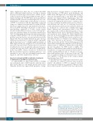
Figure 5. Communication at the endoplasmic reticu- lum-mitochondria interface in cells treated with PS89 in combination with cytostatics. For details see text. PDI: protein disulfide isomerase; BAP31: B-cell receptor- associated protein 31; ER: endoplasmic reticulum; ROS: reactive oxygen species; ΔΨm: mitochondrial mem- brane potential; PARP: poly (ADP-ribosome) polymerase.
552
haematologica | 2019; 104(3)
